1874
A comparative study of arterial spin labeling and CT perfusion on evaluation of cerebral perfusion changes after carotid endarterectomy1Radiology, Peking University Third Hospital, Beijing, China
Synopsis
3D arterial spin labeling (3D ASL) and CT perfusion (CTP) can evaluate the changes of cerebral blood flow(CBF) after carotid endarterectomy(CEA). The aim of this study is to evaluate the changes of CBF after CEA using 3D ASL and CTP respectively, and to compare the consistency of the two methods. Compared with CTP, changes of CBF values obtained by ASL were similar. ASL has similar evaluation results with CTP. As ASL is a noninvasive imaging tool, it has potential to quantitative evaluate hemodynamic changes after CEA.
Introduction
Carotid plaques cause carotid stenosis and lead to hypoperfusion of cerebral blood flow (CBF). When CBF reduced to the critical state, hypoperfusion cerebral infarction may occur. Carotid endarterectomy (CEA) is an effective surgical option for treatment of stenosis by removing plaques. There are some methods to measure CBF values, including arterial spin labeling (ASL) and CT perfusion (CTP). ASL and CTP can be used to measure CBF values and perform a follow up. The aim of this study is to evaluate the changes of CBF after CEA using 3D ASL and CTP respectively, and to compare the consistency of the two methods.Materials and Methods
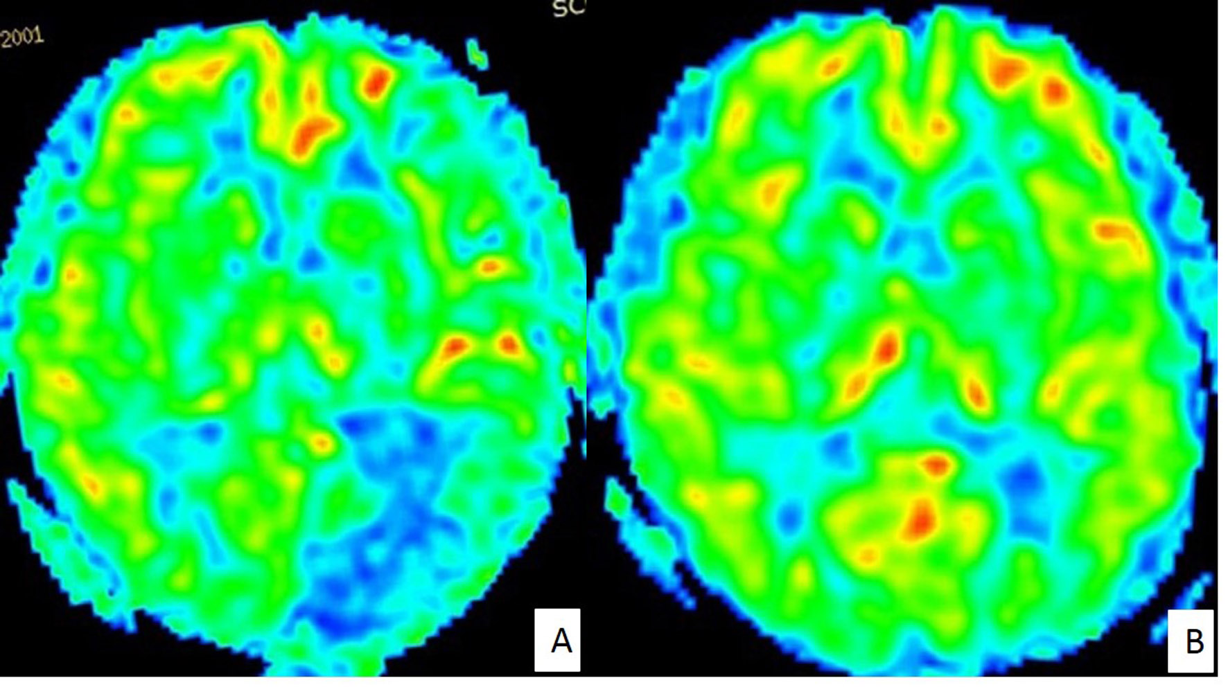
Twelve patients diagnosed as carotid artery stenosis and scheduled for CEA were recruited (mean age 67.5±4.37y; 8males, 4 females). CTP was performed with a 64 CT-MDCT and MR imaging was performed with a 3.0T MR system about 1 week before and after CEA respectively. 3D ASL acquisition parameters were: TR/TE = 4632/10.5 ms, voxel size = 2X2X4 mm3, post-labeling delay = 2000 ms. CTP acquisition parameters were: 80kV/265mA. CBF maps from 3D ASL and CTP were obtained. CBF values from ASL(Figure 1) and CTP were measured respectively in the same position of affected side and the other side before and after surgery. To assess the changes of CBF after CEA, the following differences of affected side and the other side were calculated respectively: ΔCBF=(CBFpostoperative-CBFpreoperative)/ CBFpreoperative. The ΔCBF from ASL was compared with ΔCBF from CTP.Results
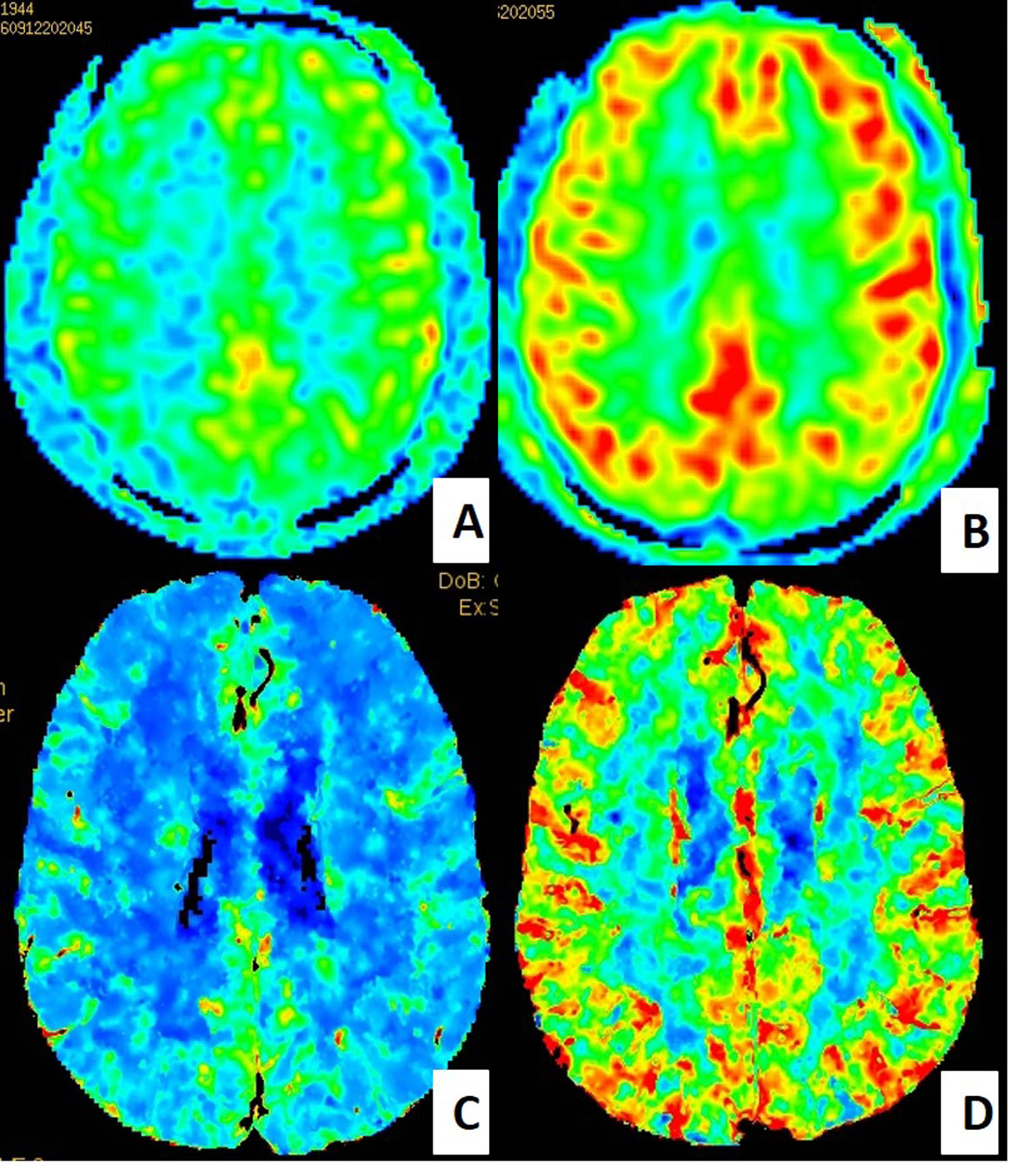
1. Compared with CBF values obtained from ASL before CEA, CBF values obtained from ASL increased after CEA, average ΔCBF of affected side was 0.280±0.345, average ΔCBF of the other side was 0.281±0.288 (Figure 2).
2. Compared with CBF values obtained from CTP before CEA, the CBF values increased after CEA: average ΔCBF of affected side was 0.119±0.190, average ΔCBF of the other side was 0.083±0.115 (Figure 2).
3. There was no significant difference of ΔCBF from 3D ASL and from CTP on effected side (P=0.340).
4. There was no significant difference of ΔCBF from 3D ASL and from CTP on the other side (P=0.164).
Conclusions
CEA improves cerebral perfusion in patients with carotid artery stenosis, which can be obtained from both ASL and CTP. Compared with CTP, changes of CBF values obtained by ASL were similar. As ASL is a noninvasive imaging tool, it has potential to quantitative evaluate hemodynamic changes after CEA.Acknowledgements
No.References
1. Nezamzadeh M, Matson GB, Young K, et al. Improved pseudo-continuous arterial spin labeling for mapping brain perfusion. J Magn Reson Imaging, 2010, 31(6):1419-1427.
2. Bokkers RP, van Laar PJ, van de Ven KC, et al. Arterial spin-labeling MR imaging measurements of timing parameters in patients with a carotid artery occlusion. AJNR Am J Neuroradiol, 2008,29(9):1698-1703.
3. Hartkamp NS, Petersen ET, De Vis JB, et al. Mapping of cerebral perfusion territories using territorial arterial spin labeling: techniques and clinical application. NMR Biomed, 2013, 26(8): 901–912.
Figures