1871
A rapid scan for simultaneous MRAV, MRA, tSWI, and QSM on 1.5T1Neusoft Medical System, Shanghai, China, 2Shanghai Key Laboratory of Magnetic Resonance, East China Normal University, Shanghai, China, 3Department of Radiology, School of Medicine, Wayne State University, Detroit, FL, United States, 4The MRI Institute for Biomedical Research, Detroit, FL, United States
Synopsis
Numerous diseases such as stroke, arteriovenous malformation (AVM), traumatic brain injury (TBI) and tumor evaluation require detailed vascular information for the best diagnostic interpretation1-4. Being able to collect both MR angiography and venography with sufficient SNR, CNR and co-registration in short time is critical for these diseases, especially for emergency patients. In this work, we developed a rapid 3D interleaved GRE sequence to acquire these vascular images simultaneously. Co-registered MRAV, MRA, QSM and tSWI for imaging arteries, veins and basal ganglia in 4 minutes and 24 seconds on a NMS 1.5T system covering the whole brain with 0.67×1.33 × 2.7 mm3 resolution
INTRODUCTION
Multiple vascular images are often necessary for neuroscientific studies and clinical applications. Conventional individual acquisition scheme takes long acquisition time for a full set of MRA, MRV, SWI and QSM. Recently, a rapid interleaved sequence for simultaneous MRAV, MRA, SWI and QSM was published which provides full brain vascular imaging in 4 minutes at 3T5. The purpose of this work is to further optimize and extend the concept to 1.5T. Co-registered MRAV, MRA, tSWI and QSM for imaging arteries, veins , basal ganglia and susceptibility map in 4 minutes and 24 seconds on a NMS 1.5T system covering the whole brain with 0.67×1.33×2.7 mm3 resolution.
METHODS
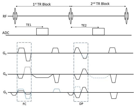
Sequence Design Similar to Ref 5, a fully flow compensation 3D GRE sequence with two TR blocks is acquired in an interleaved manner. Unlike Ref 5, there is only one echo in each TR. In the first TR block, flowing spins are rephased which providing a bright blood magnitude image. And in the second TR block flowing spins are dephased which providing a black blood magnitude image. The flow dephasing gradients are accomplished by bipolar gradients with very low velocity encoding (VENC) values. Figure 1 shows the interleaved GRE sequence.
Data Acquisition. Three healthy volunteers were scanned on a NMS S15P 1.5T system (Neusoft Medical System, Shenyang, China) with an 8-channel head coil (NMS, Shenyang, China). Using a SENSE factor of 1.5 along the L-R direction. Imaging parameters were: TR=22.5 ms, FA=15°, TE1/TE2=15 ms/15 ms, BW1/BW2=240/240Hz/pixel, voxel size=0.67 (readout) × 1.33 (phase) × 2.7 (partition) mm3, 48 transversal slices and TA = 4:24. The VENC value is about 2.5 cm/s to dephase both arteries and veins.
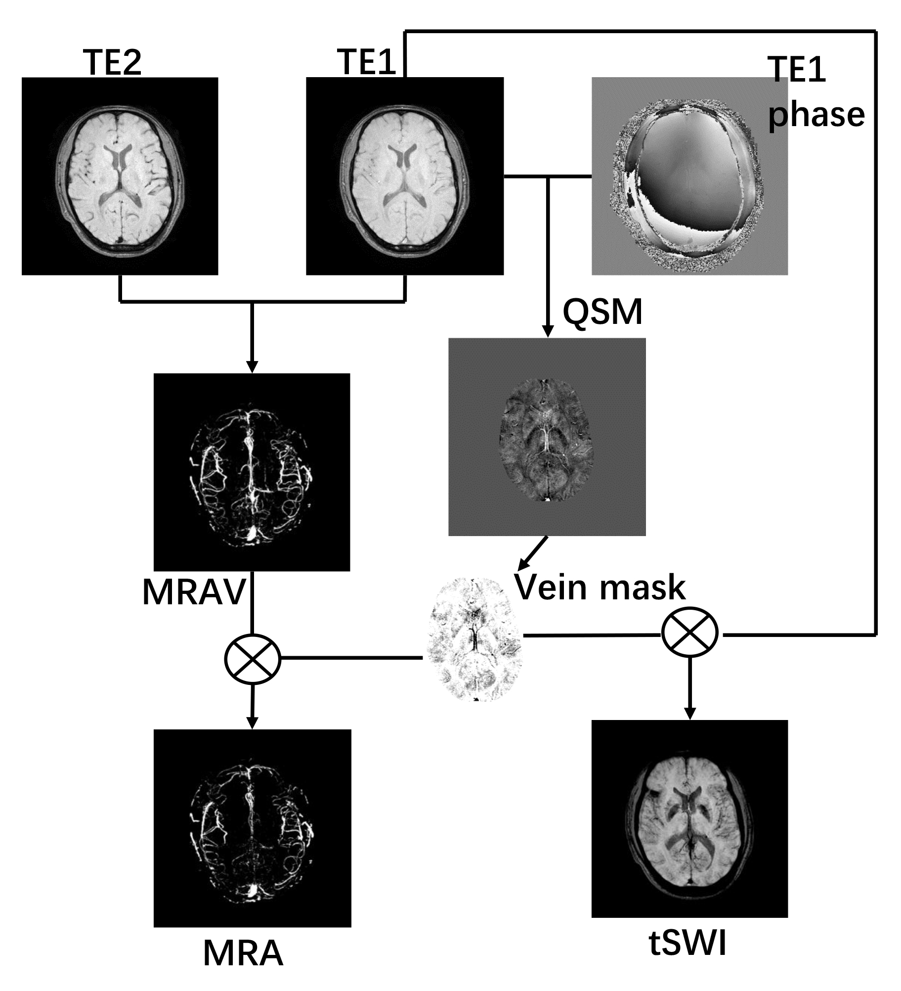
Data Processing. MRAV: The subtraction of two echoes that had identical imaging conditions but the phase preparation gave a MRAV image denoting flowing spins. QSM: iSWIM6 is used with the rephase echo data to reconstruct QSM; tSWI: The QSM map was used to generate a susceptibility mask, and then multiply it with the rephase echo magnitude image to create true-SWI image. MRA: the susceptibility mask that denoting veins was used to suppress veins on the MRAV image to generate a pure MRA image. An overview of the data processing steps is shown in Figure 2.
RESULTS
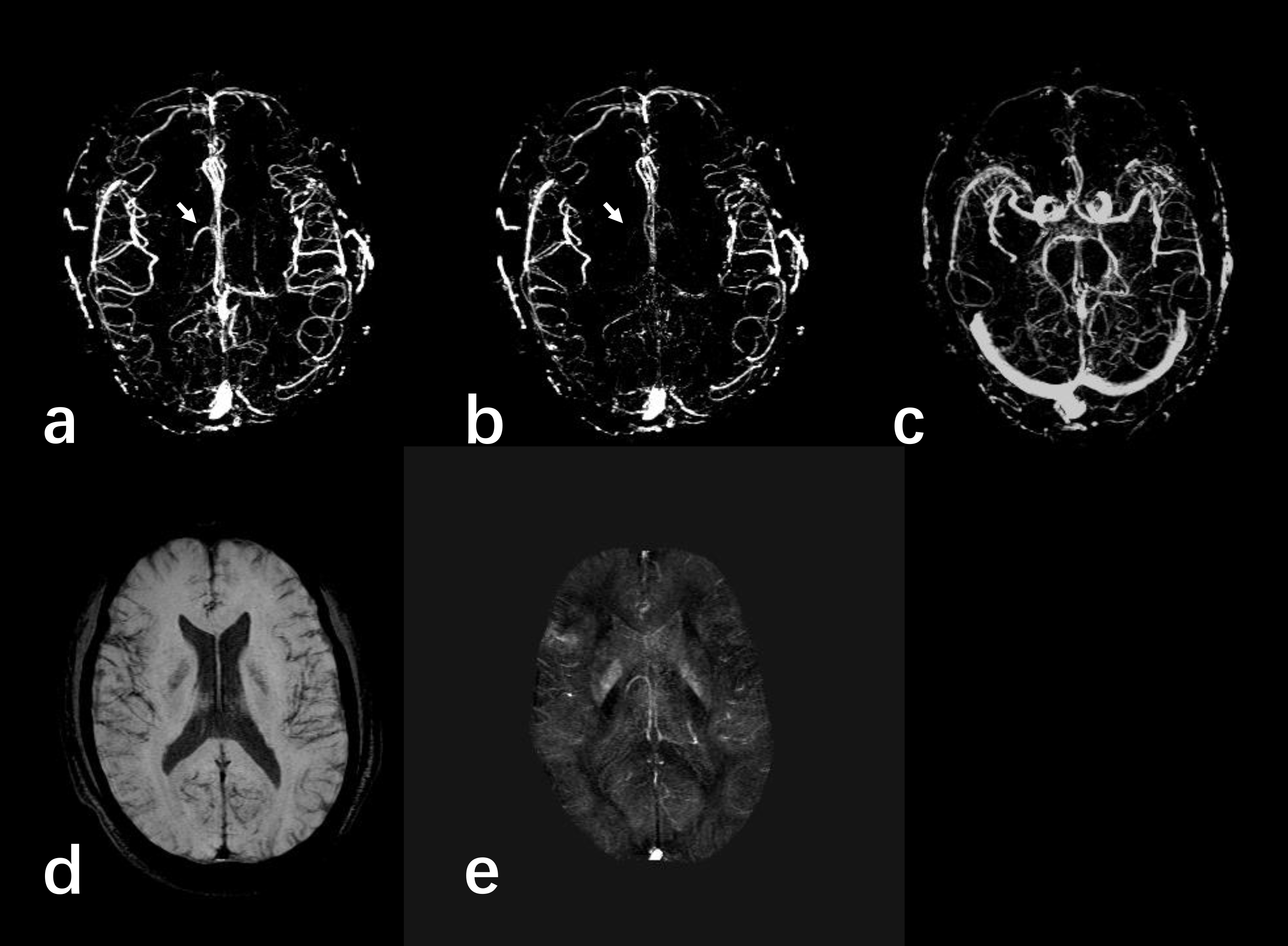
Figure 3 presents the results of one subject. MRAV, MRA, tSWI and QSM were calculated from the 2-echo data set. The MRAV showed both arteries and veins with clear background from stationary tissues. On the MRA, veins were suppressed using mask generated from QSM. Even though, the echo time is relatively shorter than conventional TE for SWI but the contrast of tSWI is still sufficient. The QSM showed nice visualization and accuracy on veins and basal ganglia.DISCUSSION
The proposed scheme herein provides one solution for acquiring high resolution and co-registered MRAV, MRA tSWI and QSM images in single less than 5-min scan. Clinically, these images can be used for studying vasculature related diseases, such as TBI and detecting thrombus and hemorrhage in stroke, tumor. Compared to the conventional individual acquisition scheme, the acquisition time for collecting the full set of the vascular data is much shorter, and all acquired images are co-registered. Moreover, the contrast between MRAV/MRA and background tissue is much better than those by conventional time-of-flight scheme; Different from Ref 5, the proposed sequence uses only one echo in the first TR, which leads to lower level requirement to acquisition system, shorter gradient duty cycle, and lower acoustic noise level.CONCLUSION
The proposed method in this work provides a clinically applicable scheme to acquire 3D MRAV, MRA, tSWI and QSM with sufficient SNR, CNR, resolution, and co-registration in single rapid scan on 1.5T.Acknowledgements
No acknowledgement found.References
[1] S Mittal, et al. American journal of neuroradiology 2009; 30:232-252. [2] EM Haacke, et al. American journal of neuroradiology 2009; 30:19-30. [3] EM Haacke, et al. Magn Reson Imaging.2015; 33:1-25. [4]Liu S, et al. NMR in Biomedicine.2016. [5] Chen Y, et al. ISMRM 2017; p1215. [6] Tang J, et al. Magn Reson Med 2013; 69(5):1396-1407.Figures