1847
Magnetic Resonance Imaging (MRI) Assessment of Dimethyl Fumarate in Protecting Myelin in a Cuprizone Mouse Model1Research and Early Development Biomarker, Biogen, Cambridge, MA, United States, 2Neuroimmunology and Acute Neurology Research Unit, Biogen, Cambridge, MA, United States
Synopsis
Multiple sclerosis (MS) is a debilitating disease that affects the central nervous system. Immune system destroys the myelin that protects the axon which leads to physical, neurocognitive, and psychiatric disorders. Symptoms may improve, but permanent neurological problems often remain. There is no known cure for MS but current treatments can improve symptoms and prevent relapse. MRI has a role in MS diagnosis and management. We demonstrated that advances in MRI techniques such as Magnetization Transfer Ratio Imaging and Diffusion Tensor Imaging can detect the protective effects of dimethyl fumarate, clinically approved MS treatment, in the corpus callosum of mice.
Introduction:
Magnetic Resonance Imaging (MRI) is currently used for the detection of multiple sclerosis (MS) lesions. Newly appearing hyperintense areas on T2-weighted (T2w) images with chronic hypointensities on T1-weighted images are used to identify areas of active MS lesions and tissue damage1. However, these MRI techniques do not measure the destructive aspects of MS pathology. Magnetization Transfer Ratio (MTR) imaging2 and Diffusion Tensor Imaging (DTI)3 have been shown to correlate significantly with myelin content. Given that these MRI methods can quantify changes related to myelin, they can be used to evaluate and screen for MS therapies. Herein, we demonstrate the utility of MTR and DTI in evaluating dimethyl fumarate (DMF), a clinically approved treatment for MS, for its protective role in the corpus callosum (CC) of the Cuprizone mouse model, which begins with cell death of oligodendrocytes followed by extensive demyelination.Methods:
All
animal handling procedures were approved by the local Institutional Animal Care
and Use Committees. C57BL/6 mice were divided into three groups
and imaged at baseline and six weeks using MTR and DTI. The groups were
divided into Wild-type-vehicle (WT-veh) treated, Cuprizone/Rapamycin-vehicle
(CR-veh) treated, and Cuprizone/Rapamycin-DMF (CR-DMF) treated. At 8 weeks, C57BL/6 mice were fed Cuprizone
that was formulated in chow (0.3%) and administered with daily Rapamycin (10
mg/kg) injections for 42 days. DMF (100
mg/kg) or vehicle was administered daily in randomized animals. MRI measurements were
performed at baseline and 6 weeks on a 7T Bruker Biospec with Paravision 5.1. Animals were
anesthetized with isoflurane carried by O2. respiration and body temperature were
monitored using the small animal monitoring system and maintained at
physiologically normal levels. A mouse
brain receive-only phased array surface coil was placed over the animal’s head
for signal reception and an 86 mm actively detuned transmit-only volume coil
was used for transmission. Imaging Sequences: High-resolution T2w Rapid Acquisition with
Refocused Echoes (RARE) were acquired with the following parameters: TR=2.6 s,
TE(eff)=83 ms, RARE Factor=16, FOV=1.6x1.67 cm, resolution=101x139x7500 um3,
slices=11, averages=10. The unsaturated
MTR was acquired using Fast Low Angle Shot (FLASH) with the following
parameters: TR=255 ms, TE=6 ms, flip angle=10o, FOV=1.61x1.67 cm,
resolution=101x104x750 um3, slices=11, averages=40. A second MTR was acquired with MT saturation
pulse (Gaussian, 10.25 ms, 10 uT, 6000 Hz off-resonance) with the same
parameters for the unsaturated. The DTI
was acquired using a EPI-Spin Echoes with the following parameters: TR=3.75 s, TE=29 ms, FOV=1.6x1.67 cm, resolution=101x139x7500
um3, slices=11, averages=3, direction=3, b-values=1200 s/mm2,
Δ=9.5 ms, δ=4.5 ms. Histopathology: 30 um
sections were stained with Black Gold for myelin. Results/Discussion:
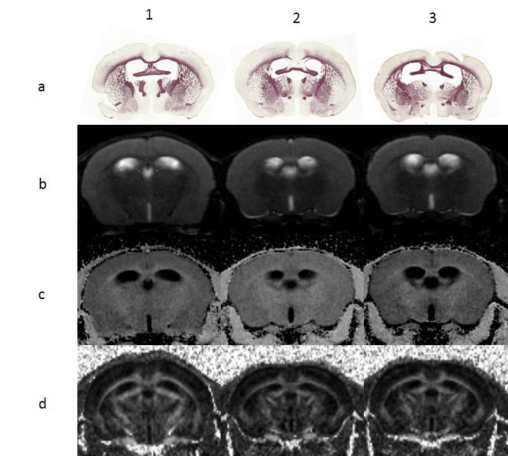
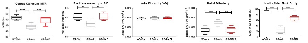
Black Gold stain and T2w image confirmed demyelination in the CC of Cuprizone demyelination model after six weeks on Cuprizone (Figure 1). At week 0, there were no significant differences between Wild-type-vehicle treated (WT-Veh), Cuprizone/Rapamycin-vehicle treated (CR-Veh), and Cuprizone/Rapamycin-DMF treated (CR-DMF) for MTR and the 3 DTI metrics: fractional anisotropy (FA), axial diffusivity (AD), and radial diffusivity (RD). FA describes the anisotropy of a diffusion process which reflects fiber density, axonal diameter, and myelination in white matter, which would be reduced in demyelination. Reduction of AD has been associated with axonal damage in white matter. Increase of RD has been associated with demyelination. At week 6, MTR, FA, and RD demonstrated significant differences in the center of CC between WT-Veh and CR-Veh; and CR-DMF and CR-Veh (Figures 1c, 1d and Figure 2). Decrease in MTR suggested a smaller macromolecular pool due to demyelination. CR-Veh showed a significant decrease in MTR while CR-DMF had a slight decrease when both were compared to WT-Veh (Figure 2). There was a significant difference between CR-Veh and CR-DMF. Next, a significant decrease in FA in CR-Veh compared to WT-Veh and CR-DMF was seen but the difference between WT-Veh and CR-DMF was small. We did not detect a significant difference in AD between the three groups. There was a significant increase in RD in CR-Veh compared to WT-Veh and DR-DMF, but no difference between WT-Veh and CR-DMF. There was a significant decrease in the myelin area in the Black Gold stained mid-CC in CR-Veh compared to WT-Veh and CR-DMF. These findings all suggest that there is a protective effect of DMF against demyelination effects of Cuprizone.Conclusion:
We demonstrated the ability of MTR and DTI to detect the cytoprotective and immunomodulatory effects of DMF in the corpus callosum. These two methods can be used in conjunction with other modalities as quantitative methods for measuring myelin in the CNS and can serve as a tool to evaluate and screen for potential MS therapies.Acknowledgements
No acknowledgement found.References
1. Rovia A, Auger C, Alonso J. Magnetic resonance monitoring of lesion evolution in multiple sclerosis. Ther Adv Neurol Disord. 2013;6(5):298-310
2. Schmierer K, Scaravilli F, Altmann DR, et al. Magnetization transfer ratio and myelin in postmortem multiple sclerosis brain. Ann Neurol. 2005;56(3):407-15
3. Song SK, Yoshino J, Lin SJ, et al. Demyelination increases radial diffusivity in corpus callosum of mouse brain. Neuroimage. 2005;26(1):132-40
Figures