1845
Recuperative white matter integrity in long-term abstinent heroin addicts1Radiology, Tangdu Hospital, The Fourth Military Medical University, Xi’an, China
Synopsis
Heroin-induced white matter integrity disruption and the restorability during long-term abstinence have been reported. However, the characteristic of these recover during different stage of abstinence has not been well understood. Use the voxel-wised diffusion tensor method,we compared the white matter difference within 17 long-term abstinence heroin addicts (LA), 22 short-term abstainers (SA) and 20 healthy controls (HC). We found significantly decreased white matter integrity in SA and the time-dependent recover of white matter integrity, especially the restoration of myelin sheath, in LA,. These structural recover may contributed to the improvement of function in the duration of long-term abstinence.
Purpose
Heroin addiction is a serious social problem in China. Neuroimaging and cognitive test have demonstrated that heroin-related structural and cognitive impairment would have the tendency to recover during long-term abstinence[1-3]. However, the influence of abstinence to brain white matter and the characteristic of these effects during different stage of abstinence has not been well understood. The aim of the present study was to explore white matter characteristics of heroin addicts in short-term and long-term abstinence by a quantitative DTI method.Materials and Methods
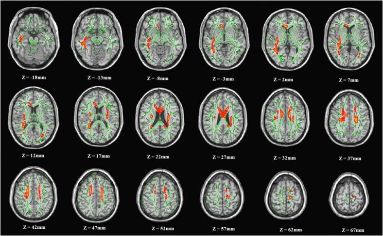
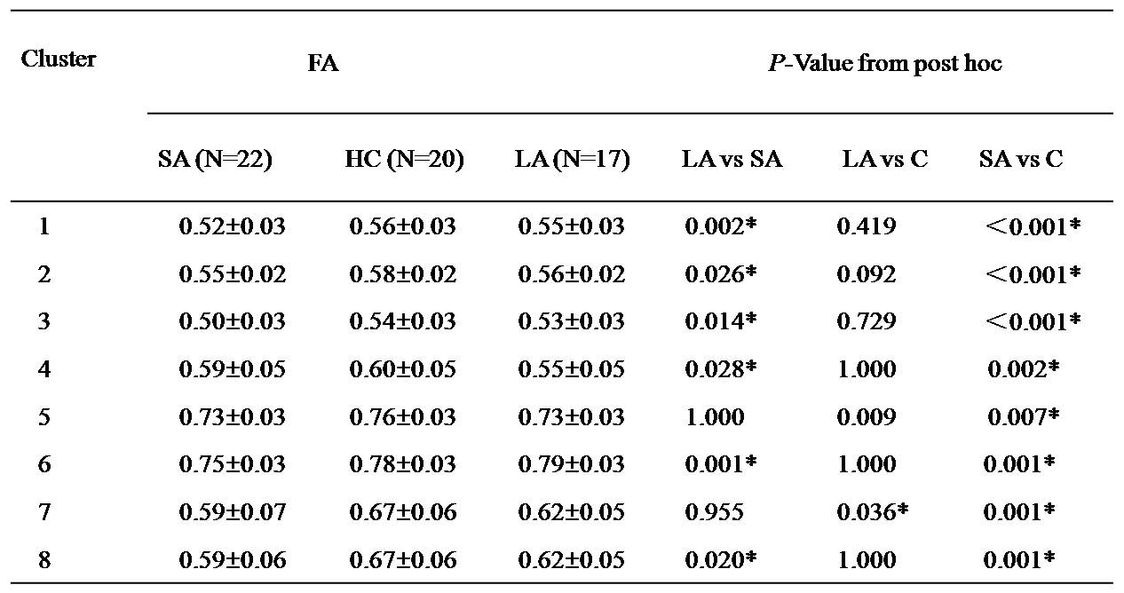
Seventeen long-term abstinence heroin addicts (LA), 22 short-term abstainers (SA) and 20 healthy controls (HC) participated in this study. Diffusion tensor imaging data were acquired on 3.0T GE Signa Excite HD whole-body MRI system. FMRIB's Diffusion Toolbox (FDT, http://fsl.fmrib.ox.ac.uk/fsl/fslwiki/FDT) and Tract-based spatial statistics (TBSS) from FMRIB's Software Library (FSL, version 4.1.8, http://fsl.fmrib.ox.ac.uk/fsl/fslwiki/) were used for data preprocessing and white matter microstructure analysis. Design of ANOVA were used for comparison of fractional anisotropy (FA). The radial diffusivity (RD) and axial diffusivity (AD) were inducted to evaluate the source of the abnormal FA change. The correlation between DTI indices and the duration of abstinence were calculated using Pearson correlation.Results
Compared with HC, SA had significantly reduced FA in extensive WM regions, such as corticospinal tract , corona radiata , superior longitudinal fasciculus , external capsule , corpus callosum. While LA had only two regions showed the significant reduction of FA, located in left inferior fronto-occipital fasciculus and superior longitudinal fasciculus. When compared to SA, LA had significantly increased FA. The alteration of FA were detected caused by the increase of the RD, while no significant difference occurred in AD. In all abstinent patients, FA values in most of the differential regions were correlated positively with the duration of the abstinence, while the RD values in these regions were correlated negatively with the duration of the abstinence.Discussion and Conclusion
These findings suggested that the time-dependent recover of white matter integrity, especially the restoration of myelin sheath, were occurred in long-term abstinence. These structural recover may contributed to the improvement of function in the duration of long-term abstinence.Acknowledgements
This work was supported by the grants from the National Natural Science Foundation of China (Nos. 81401393 and 81371532).References
1. Li, W., et al., White matter impairment in chronic heroin dependence: A quantitative DTI study. Brain Res, 2013. 1531: p. 58-64.
2. Morie, K.P., et al., Intact inhibitory control processes in abstinent drug abusers (II): A high-density electrical mapping study in former cocaine and heroin addicts. Neuropharmacology, 2013.
3. Wang, X., et al., Reversible brain white matter microstructure changes in heroin addicts: a
Figures