1844
Brain Gray Matter Abnormalities in First-Episode, Treatment-Naïve Patients with Obsessive-Compulsive Disorder1The First Affiliated Hospital of Zhengzhou University, Zhengzhou City, China, 2GE Healthcare, MR Research China, Zhengzhou, China, 3The First Affiliated Hospital of Zhengzhou University, Zhengzhou, China
Synopsis
Examinations of 36 first-episode, treatment-naive pediatric OCD patients without any comorbidities and 37 matched healthy controls (HCs) were performed with 3.0T magnetic resonance imaging (MRI). Voxel-based morphometry (VBM) following Diffeomorphic Anatomical Registration using Exponentiated Lie algebra (DARTEL) was used to conduct voxel-wise tests for group differences in regional gray matter volume (GMV). Compared to HCs, the patient group exhibited significantly different GMV in bilateral anterior cingulate cortex (ACC), left fusiform gyrus and the left postcentral gyrus. It is believed that this noninvasive method might be useful for exploring the pathophysiology of OCD.
Purpose
Obsessive-compulsive disorder (OCD) is a psychological disorder with an estimated lifetime prevalence of 1% to 3% 1, characterized by uncontrollable obsessive thoughts, compulsive behavior and compulsions to counteract these thoughts and behavior clinically 2, which is among the leading causes of functional disability worldwide, but the neurobiology and aetiological origins of it is not clear. Recently, there is increased evidence of widespread structural alterations in gray matter in OCD patients 3, so that the objective of this study was to test the existence of structural alterations in gray matter between OCD patients and healthy control groups using voxel-based morphometry (VBM) method.Method
A total of 73 subjects were recruited including 36 drug-naive OCD patients and 37 healthy controls with matched age, gender, handedness and education level in this study (As Table1 showed). OCD patients were diagnosed through Structured Clinical Interview for DSM-V Axis I Disorders-Non-patient Edition (SCID-I) by an experienced psychiatric chief physician. MRI images of all subjects were acquired using a 3.0 Tesla MR scanner (DiscoveryMR750, General Electric, Milwaukee, WI, USA). Sagittal 3D T1-weighted images were acquired by a brain volume sequence with the following parameters: repetition time/echo time=8.2/3.2ms; field of view=256×256mm2; matrix=256×256; slice thickness=1.0 mm, no gap; 188 slices. Whole brain GMV was calculated using the optimized VBM technique implemented within SPM8. Firstly, the 3D-T1WI structure images were segmented into gray matter (GM), white matter and cerebrospinal fluid. And then segmented images were registered into a strictly aligned space. Subsequently, the non-linear warping of GM images was performed using exponentiated Lie algebra (DARTEL) technique. Finally, demographics and clinical variables were assessed using statistical software (SPSS, version 17.0). Meanwhile, the differences in GMV between two groups were analyzed in VBM-DARTEL using an independent samples t-test with whole brain volume, age, gender, and education as covariates. Note that statistical maps were created using a combined threshold of P <0.001 and a minimum cluster size of 100 voxels.Results
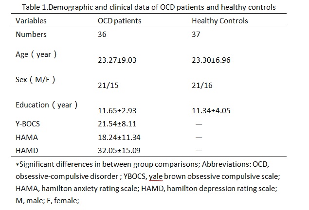
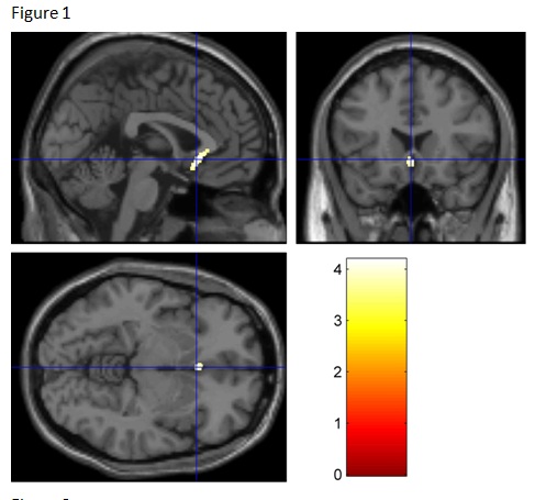
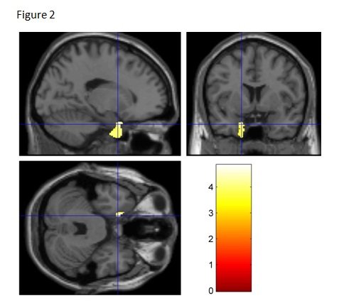
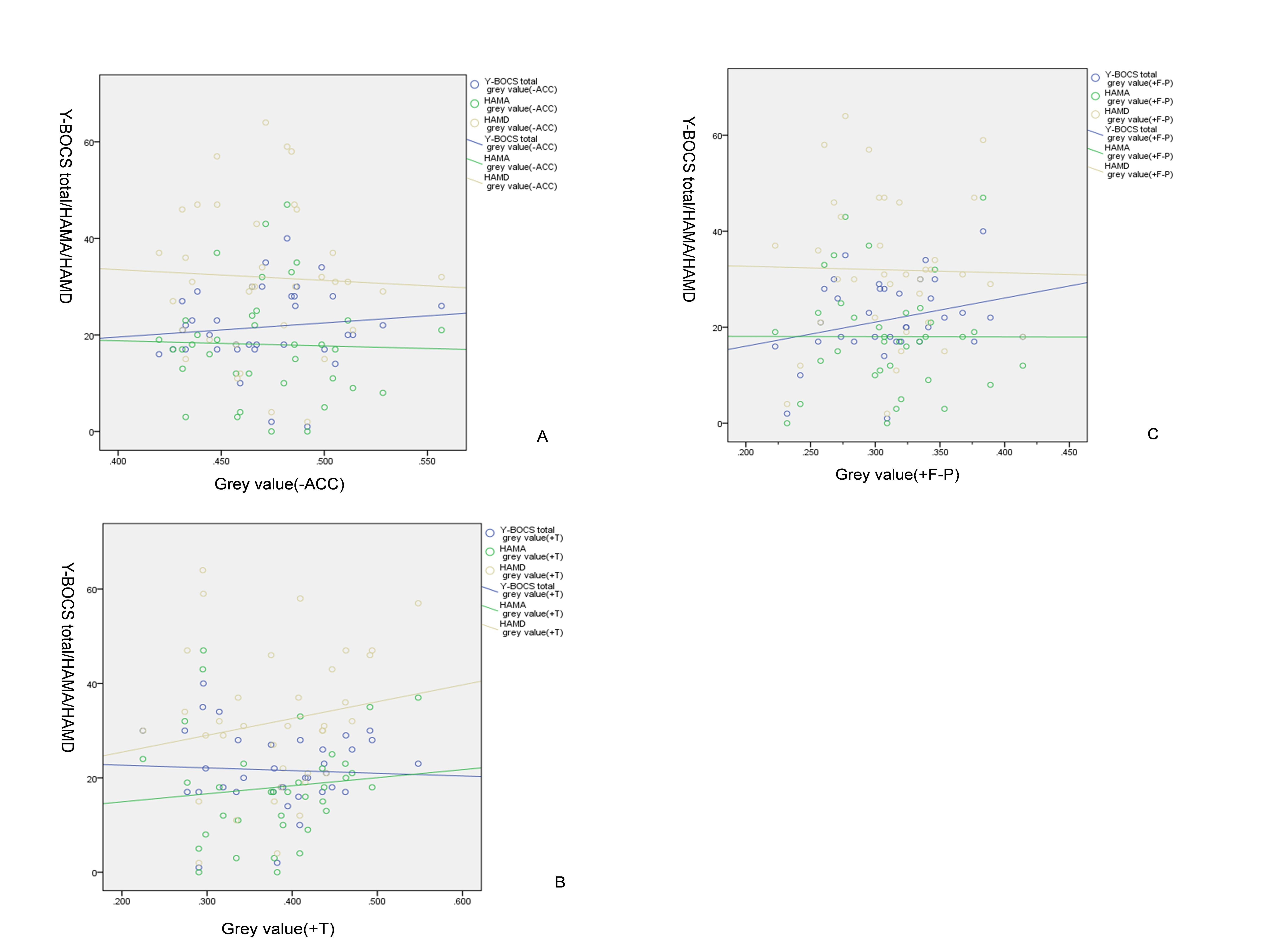
In this study, the results suggest that the OCD patients group exhibited significantly less GMV in bilateral anterior cingulate cortex (ACC) (T=-4.181, P=0.0005) and significantly more GMV in the left fusiform gyrus (T=4.841, P=0.0005) and the left postcentral gyrus (T=4.299,P=0.0005) than healthy controls group as indicated in Figure 1, 2, and 3 respectively. In addition, the GMV alternation had no correlation with Yale brown obsessive compulsive scale (YBOCS), Hamilton anxiety rating scale (HAMA) and Hamilton depression rating scale (HAMD) scores as indicated in Figure 4.Discussion
The results of whole brain gray matter volume (GMV) analysis in OCD patients without drug and psychosocial interventions offered more objective and reliable information for psychological mechanism of OCD. Most of structural MRI studies on OCD patients were based on volumetric region of interest (ROI) methods, and few studies utilized voxel-based morphometry (VBM). Besides, DARTEL was found to rank the top level to optimize the sensitivity of such analysis and detect subtle brain structural changes than those measured with standard VBM. Our current results suggest that the GM abnormalities were defined in the OCD patients. Moreover, these findings provided further evidence of brain GM abnormalities that are not only present in the classical cortical–striatal–thalamic–cortical (CSTC) circuit but also besides the CSTC circuit, which may represent the complex interaction of abnormally functional organization of networks in OCD.Conclusion
In our study, the differences in GMV between two groups were assessed in VBM-DARTEL. OCD patients showed significantly decreased GMV in the bilateral anterior cingulate cortex and increased GMV in the left fusiform gyrus and the left postcentral gyrus compared to HCs, which may represent the complex interaction of abnormally functional organization of networks in OCD. It is believed that this noninvasive method might be useful for exploring the pathophysiology of OCD.Acknowledgements
No acknowledgement found.References
[1] Ruscio AM, et al. (2010). The epidemiology of obsessive—compulsive disorder in the National Comorbidity Survey Replicatio. Mol Psychiatry. 15:53-63.
[2] Burdick KE, et al. (2008). Neurocognitive profile analysis in obsessive compulsive disorder. J Int Neuropsychol Soc. 14:640–645.
[3] Menzies L, et al. (2008). Integrating evidence from neuroimaging and neuropsychological studies of obsessive-compulsive disorder: the orbitofronto-striatal model revisited. Neurosci Biobehav Rev. 32:525-549.
Figures