1841
Gray Matter Network Organization in Psychotic DisordersWenjing Zhang1, Du Lei1, Brett Clementz2, Carol Tamminga3, Matcheri Keshavan4, Sarah Keedy5, Godfrey Pearlson6, Elliot Gershon5, Jeffrey Bishop7, Jieke Liu1, Qiyong Gong1, John Sweeney8, and Su Lui1
1Huaxi MR Research Center (HMRRC), Department of Radiology, West China Hospital of Sichuan University, Chengdu, China, 2Department of Psychology, University of Georgia, Athens, GA, United States, 3Department of Psychiatry, University of Texas Southwestern Medical Center, Dallas, TX, United States, 4Department of Psychiatry, Beth Israel Deaconess Medical Center, Harvard Medical School, Boston, MA, United States, 5Department of Psychiatry and Behavioral Neuroscience, University of Chicago, Chicago, IL, United States, 6Department of Psychiatry, School of Medicine, Yale University, New Haven, CT, United States, 7Department of Experimental and Clinical Pharmacology, College of Pharmacy, University of Minnesota, Minneapolis, MN, United States, 8Department of Psychiatry, University of Cincinnati, Cincinnati, OH, United States
Synopsis
Recently, new approaches have been developed using graph theory to identify deficits in gray matter networks at individual level. In the current study, by investigating single-subject graphs based on gray matter morphology to define neuroanatomic networks in a large group of individuals across psychotic disorders (n=330), we observed disrupted network organizations associated with superior temporal and prefrontal regions within the gray matter networks in patients, which were also negatively associated with severity of psychotic symptoms. These findings showed the utility of graph theory based measures of neuroanatomic network organization to extend our understanding of the neurobiology underlying psychotic disorders.
INTRODUCTION
Recent studies have identified discrete clusters of patients across the spectrum of psychotic disorders with defining characteristics including features of gray and white matter, 1 regional brain activity 2 and resting-state connectivity.3 Many of these studies focused on regional brain deficits while the neuropathology of psychotic disorders is believed to be more closely to abnormal network organization. 4 Previous studies have typically characterized network organization via fMRI measures or write-matter connectivity measures. However, the functional changes depend greatly on the brain states whereas the white-matter measures could be easily affected by tractography algorithms and the measures used for the graph construction. Recently, new approaches have been developed using graph theory to identify deficits in gray matter networks that have now been developed for use in individual patients.5 In the present study, we aimed to demonstrate the feasibility and utility of this new approach for identifying atypical gray matter network alterations in a large cohort of patients with serious mental illness.METHODS
N=854 total participants from the B-SNIP-1 consortium were included in this study, including 330 psychosis probands (109 with schizophrenia, 88 with schizoaffective disorder, and 133 with psychotic bipolar disorder), plus 320 of their nonpsychotic first degree relatives and 204 healthy controls. Detailed descriptions of how the B-SNIP clinical population was enrolled and clinically assessed are available. 6 We followed the methodology proposed by Tijms et al 5 to extract individual structural morphology brain networks, and extended it according to Batalle et al 7 to normalize networks to a common comparable framework based on the AAL parcellation template. In gray matter graphs, nodes represent small cortical areas whereas edges represent statistical similarities in regional gray matter morphology between nodal regions. GRETNA toolbox (www.nitrc.org/projects/gretna/) was used to calculate the global and nodal network topological properties of individual brain networks. In statistical analysis, we identified differences in network organization between patients, their nonpsychotic relatives and controls with age, sex, race and handedness included as covariates. Altered structural network metrics in probands were also correlated with symptom severity.RESULTS
All probands, their nonpsychotic relatives and healthy controls showed small-world architectures (i.e., σ > 1) at all connection densities. There were no significant differences among the three participant groups in global network properties. However, significant group differences were found in regional network organization. Relative to both healthy controls and nonpsychotic relatives, probands showed decreased nodal degree in right superior frontal gyrus (SFG) and bilateral superior temporal gyrus (STG), and lower nodal efficiency in bilateral STG (p<0.05, Bonferroni corrected). There were no significant differences between nonpsychotic relatives and healthy controls in any regional network metrics (Figure 1). In probands, nodal efficiency of left and right STG was negatively associated with severity of psychotic symptoms as revealed by PANSS scores.DISCUSSION
Compared to functional MRI networks, structural networks analysis may reflect more stable patterns of anatomical organization under conditions, and provide novel information relevant to the neuropathology of brain disorders. The gray matter network organizations of psychotic probands showed decreased nodal degree and nodal efficiency mainly in superior temporal gyrus but also in superior prefrontal cortex. In previous graph theory based analyses using functional connectivity data, superior temporal regions have also been found to have reduced local connectivity in psychotic disorders. 8 Deficits of superior temporal cortex may be clinically relevant because of their potential relevance for psychotic symptomatology such as auditory hallucinations. 9 This point is consistent with our observation of an association between PANSS positive symptom scores and decreased nodal efficiency in superior temporal regions. Thus, our findings show the utility of graph theory based measures of neuroanatomic network organization for understanding the neurobiology of psychotic disorders.CONCLUSION
By investigating single-subject graphs based on gray matter morphology to define neuroanatomic networks in a large group of individuals across disorders, our findings provide novel evidence indicating gray matter disorganizations mainly involving the superior temporal regions that was related to the severity of psychotic symptomology.Acknowledgements
No acknowledgement found.References
- Ivleva EI, Bidesi AS, Keshavan MS, et al. Gray matter volume as an intermediate phenotype for psychosis: Bipolar-Schizophrenia Network on Intermediate Phenotypes (B-SNIP). Am J Psychiatry 2013; 170:1285-1296.
- Meda SA, Wang Z, Ivleva EI et al. Frequency-Specific Neural Signatures of Spontaneous Low-Frequency Resting State Fluctuations in Psychosis: Evidence From Bipolar-Schizophrenia Network on Intermediate Phenotypes (B-SNIP) Consortium. Schizophr Bull 2015; 41:1336-1348.
- Meda SA, Ruano G, Windemuth A et al. Multivariate analysis reveals genetic associations of the resting default mode network in psychotic bipolar disorder and schizophrenia. Proc Natl Acad Sci U S A 2014; 111:E2066-2075.
- Fornito A, Zalesky A, Breakspear M: The connectomics of brain disorders. Nat Rev Neurosci 2015; 16:159-172.
- Tijms BM, Series P, Willshaw DJ, Lawrie SM: Similarity-based extraction of individual networks from gray matter MRI scans. Cereb Cortex 2012; 22:1530-1541.
- Tamminga CA, Ivleva EI, Keshavan MS et al. Clinical phenotypes of psychosis in the Bipolar-Schizophrenia Network on Intermediate Phenotypes (B-SNIP). Am J Psychiatry 2013; 170:1263-1274.
- Batalle D, Munoz-Moreno E, Figueras F, Bargallo N, Eixarch E, Gratacos E: Normalization of similarity-based individual brain networks from gray matter MRI and its association with neurodevelopment in infants with intrauterine growth restriction. Neuroimage 2013; 83:901-911.
- Alexander-Bloch AF, Gogtay N, Meunier D et al. Disrupted modularity and local connectivity of brain functional networks in childhood-onset schizophrenia. Front Syst Neurosci 2010; 4:147.
- Barta PE, Pearlson GD, Powers RE et al. Auditory hallucinations and smaller superior temporal gyral volume in schizophrenia.Am J Psychiatry. 1990;147(11):1457-62.
Figures
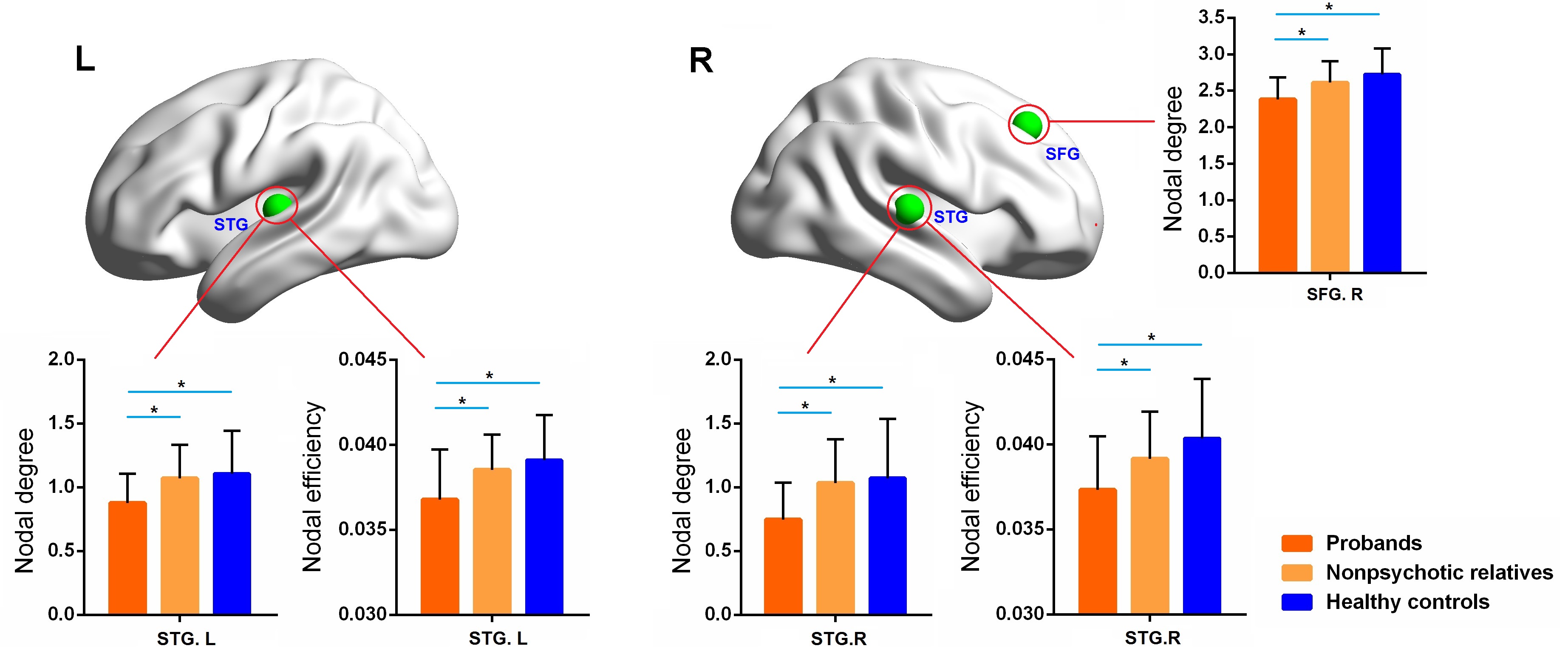
Figure 1. Nodal network metrics showing significant inter-group differences among probands, their nonpsychotic relatives andhealthy controls.