1840
White Matter Abnormalities in Never-Treated Patients with Long Term SchizophreniaYuan Xiao1, Huaiqiang Sun1, Bo Tao1, Youjin Zhao1, Wenjing Zhang1, Qiyong Gong1, John Adrian Sweeney2, and Su Lui1
1Dept. of Radiology, West China Hospital of Sichuan University, Chengdu, China, 2Dept. of Psychiatry and Behavioral Neuroscience, University of Cincinnati, Cincinnati, OH, United States
Synopsis
Do white matter abnormalities increase over the long-term course of schizophrenia, and is their trajectory influenced by antipsychotic treatment? In this cross-sectional study, more alteration of white matter microstructure were found in long-term but never-treated schizophrenia patients than duration-matched chronically treated patients. In the genu of the corpus callosum, there was an accelerated age-related reduction of fiber tract integrity in the never-treated patients. The more attenuated white matter changes in the treated patient group suggests that long-term antipsychotic treatment may have a neuroprotective effect on white matter tracts.
Introduction
Abnormalities of cerebral white matter have been reported in schizophrenia1-4. However, whether those changes are mainly due to the disorder or are secondarily to antipsychotic treatment remains unclear, as is the issue of whether they are progressive. Separating influences of these two mechanisms remains challenging because nearly all patients are appropriately treated following diagnosis, and antipsychotic drugs have robust effects on brain anatomy5-7. Comparing never-treated and antipsychotic-treated long term schizophrenia patients could shed light on white matter changes over the longer term course of illness and whether these affects are influenced by antipsychotic drugs.Methods
This study was IRB approved and written informed consent was obtained from each participant. Thirty-one never-treated, long-term schizophrenia patients with illness duration ranging from 5 to 47 years, 46 illness duration matched schizophrenia patients who had received long-term antipsychotic-treatment, and 58 healthy controls underwent DTI studies. Routine DTI preprocessing were performed using FSL software. Then, we use Automatic Fiber Quantification software to identify 20 white matter tracts (JHU white matter tractography altas) in individual subjects. Tract profiles for fractional anisotropy (FA) of white matter tracts were extracted and compared among groups using two-way (3 groups × 20 regions) ANOVAs. Significant effects were followed by post-hoc one way ANOVAs comparing groups on each tract separately, and then pairwise post hoc tests to determine which of the 3 groups differed significantly using 10000 permutation testing. Linear regression analyses were used to explore the potential relationship between FA for tracts that showed significant group differences and age among the three groups.Results
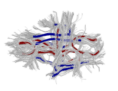
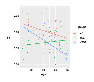
FA significantly differed among the three groups in 14 of 20 white matter tracts defined in the JHU white-matter template (P<0.05). Compared to antipsychotic-treated patients, untreated long term schizophrenia patients displayed significantly reduced FA in bilateral anterior thalamic radiation, left cingulum-hippocampus pathway, splenium and genu of corpus callosum, left superior longitudinal fasciculus and left superior longitudinal fasciculus-temporal part, and greater FA in right uncinate fasciculus (P<0.05, Figure 1). Furthermore, untreated patients showed an accelerated age-related reduction of FA in the genu of the corpus callosum relative to both treated patients and controls (P<0.05, Figure 2).Conclusion
The current study revealed more alteration of white matter microstructure in long-term never-treated schizophrenia patients than chronically treated patients of similar illness duration. In the genu of the corpus callosum, in which fibers connect left and right frontal cortex, there was an accelerated age-related reduction of fiber tract integrity in the long-term never-treated chronic schizophrenia patients not seen in treated group. These findings provide insight into disease-related white matter deficits in the years after illness onset in schizophrenia, and suggest that long-term antipsychotic treatment may have a neuroprotective effect on white matter tracts over the longer-term course of illness.Acknowledgements
This study was supported by the National Natural Science Foundation (grant numbers 81621003, 81671664 and 81371527), National Youth Top-notch Talent Support Program of China and the Program for Changjiang Scholars and Innovative Research Team in University (PCSIRT, IRT1272) of China.References
- Ellison-Wright I, & Bullmore E. Meta-analysis of diffusion tensor imaging studies in schizophrenia. Schizophr Res. 2009;108(1-3): 3-10.
- Cropley V L, Klauser P, Lenroot R K, et al. Accelerated Gray and White Matter Deterioration With Age in Schizophrenia. Am J Psychiatr. 2017;174(3):286-295.
- Kanaan R A, Kim J S, Kaufmann W E, et al. Diffusion tensor imaging in schizophrenia. Biol Psychiatry. 2005;58(12):921-929.
- Kelly S, Jahanshad N, Zalesky A, et al. Widespread white matter microstructural differences in schizophrenia across 4322 individuals: results from the ENIGMA Schizophrenia DTI Working Group. 2017;doi: 10.1038/mp.2017.170.
- Konopaske G T, Dorph-Petersen K A, Pierri, J N, et al. Effect of chronic exposure to antipsychotic medication on cell numbers in the parietal cortex of macaque monkeys. Neuropsychopharmacology. 2007;32(6):1216-1223.
- Konopaske G T, Dorph-Petersen K A, Sweet R A, et al. Effect of chronic antipsychotic exposure on astrocyte and oligodendrocyte numbers in macaque monkeys. Biol Psychiatry. 2008;63(8): 759-765.
- Keshavan M S, Bagwell W W, Haas G L, et al. Changes in caudate volume with neuroleptic treatment. Lancet. 2008;344(8934):1434.
Figures

Illustration of white matter tracts (in red color) that showed
significant difference of fractional anisotropy (FA) between never-treated
long-term schizophrenia patients and antipsychotic-treated patients of similar
illness duration.

Linear regression between age and mean FA of the genu of corpus callosum
in never-treated long-term schizophrenia patients, treated patients of similar
illness duration and healthy comparison subjects.