1836
Investigation of resting-state fMRI and cognitive function changes in patients with late-onset depression after one year follow-up1Radiology, Ruijin Hospital,Shanghai Jiao Tong University School of Medicine, Shanghai, China, 2Pudong new area mental health center, Shanghai, China, 3Ruijin Hospital, Shanghai Jiao Tong University School of Medicine, Shanghai, China
Synopsis
Late-onset depression is a common psychiatric disorder, depressed elderly often exhibit cognitive impairment that are substantial, prevalent, and disabling. The LOD patients with cognitive impairment has increased risk of conversion to dementia. The amplitude low-frequency fluctuation analysis based on resting state fMRI can directly reflect the intensity of spontaneous activity of neurons and provide information of local neurons in brain areas. In this study, we observed the changes of cognitive function and local brain functional activity in patients with LOD after one year follow-up, investigated the correlation between cognitive function and brain activity. And possibly provide an objective imaging basis for the early intervention in LOD patients with cognitive impairment before deteriorate into dementia.
Objective
To investigate the characteristics of cognitive function changes in patients with late-onset depression (LOD), and use resting-state fMRI based on amplitude of low frequency fluctuation (ALFF) method to observe the changes of local brain function in patients with LOD, and explore the correlation between cognitive function and brain activity.Methods
22 LOD patients and 11 normal controls (NC) were enrolled in the study. All subjects underwent Mini-mental State Examination (MMSE), Montreal cognitive assessment (MoCA) and complete Chinese version of neuropsychological test battery (NTB) , and the resting-state Bold-fMRI data were acquired and preprocessed. Analysis of whole brain ALFF between patients group and the control group were performed by two sample t test, to evaluate the changes of local brain functions in patients with LOD. Then correlation analysis was performed between the ALFF values of abnormal brain areas and clinical scale scores.Results
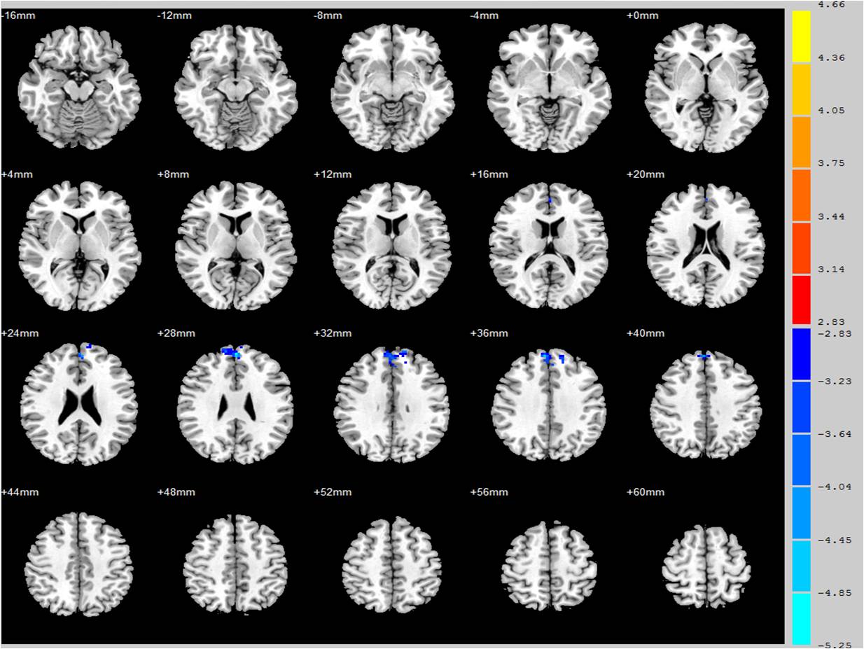
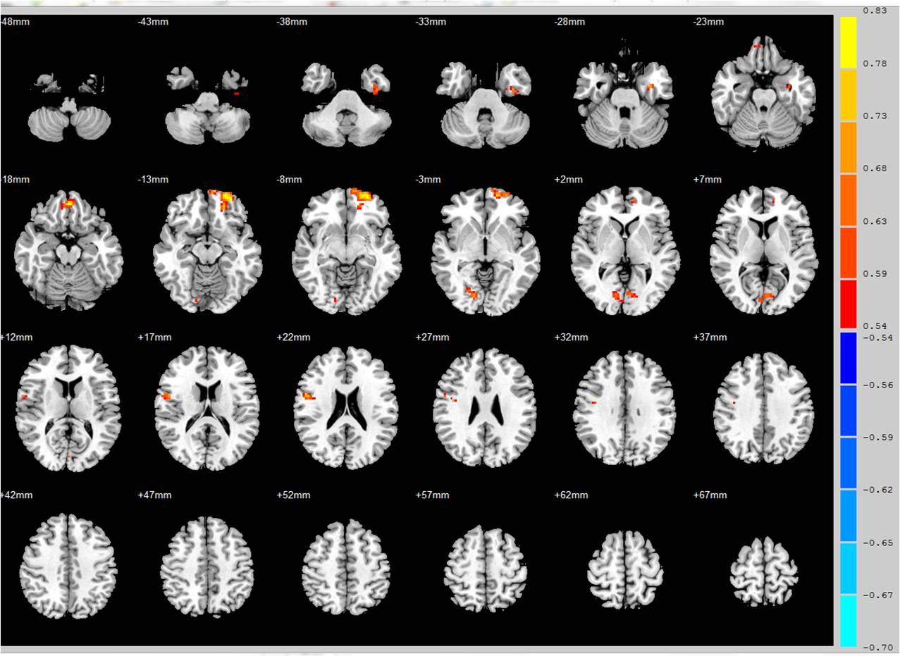
Compared with the baseline state of the patients group, the scores of MMSE delayed recall and MoCA sustained attention after one year follow-up significantly decreased, and the total score of NTB associative learning test was significantly increased (P <0.05). There was no statistically significant difference in the scores of NC group after one year follow-up.Compared with the NC group in baseline state, patients group showed significantly reduced ALFF value in left orbital frontal gyrus, left middle frontal gyrus and increased ALFF value in right middle frontal gyrus, dorsolateral prefrontal cortex; after one year follow-up, patients group showed significantly increased ALFF value in prefrontal cortex, include bilateral superior and middle frontal gyrus. Correlation analysis showed a positive correlation between the ALFF value of patients group in left prefrontal cortex/inferior temporal gyrus, bilateral occipital lobe, right precentral gyrus and the total score of NTB associative learning test.Conclusion
After one year follow –up, LOD patients exhibited cognitive impairment and showed changes of ALFF value in prefrontal cortex. The impairment of cognitive function may be the performance of abnormal activity in some brain regions. The functional changes in different brain regions, especially in prefrontal cortex, possibly related to the short-term memory indicated by NTB associative learning test.Acknowledgements
The authors thank Dr. Hongmei Fu and Dr. Hengfen Gong for the recruiting part of the volunteers. The authors thank Dr. Naying He for statistical analysis. The authors wish to thank all the subjects who participated in this study.References
No reference found.Figures
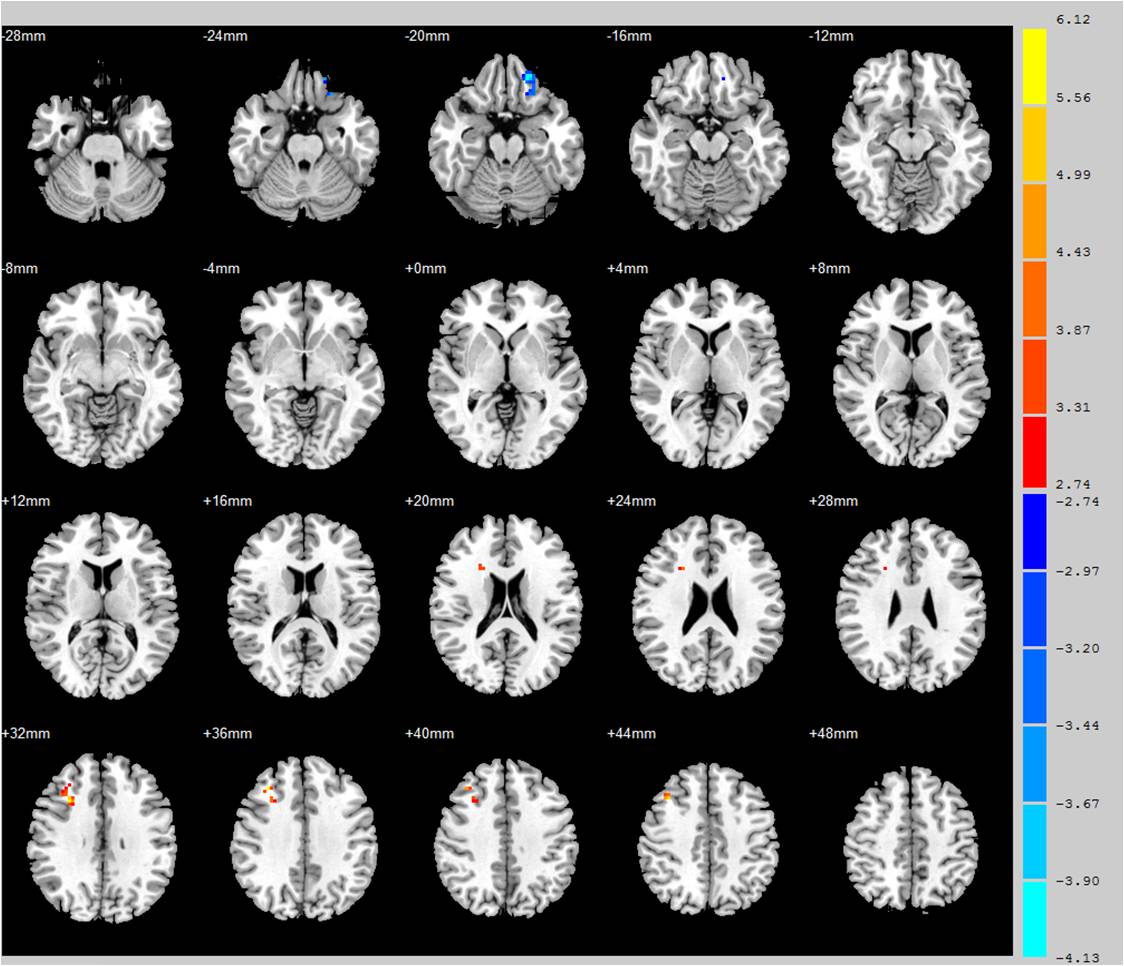
Figure 1 ALFF values with statistically significant difference showed in different brain areas when LOD group compared with NC group in baseline state.