1833
A meta-analysis of altered resting-state functional activity in medication-naive patients with first-episode major depression versus healthy controlsXiaoyue Ma1, Jia Liu2, Taiyuan Liu1, Yan Wang1, Meiyun Wang1, and Tianyi Qian3
1Zhengzhou University People’s Hospital & Henan Provincial People’s Hospital& Henan Key Laboratory for Medical Imaging of Neurological Diseases, Zhengzhou, China, 2Union Hospital, Tongji Medical College, Huazhong University of Science and Technology, Wuhan, China, 3Siemens Healthcare, MR Collaboration NE Asia, Beijing, China
Synopsis
This study aimed to use the voxel-based meta-analytic technique called anisotropic effect size-signed differential mapping (AES-SDM) to determine consistent regional brain activity alterations in medication-naive patients with first-episode unipolar major depression disorder (MDD) versus healthy controls (HCs). The pooled and subgroup meta-analyses found that MDD patients showed resting-state brain decreased activity in the left anterior lobe of the cerebellum and increased activity in the left amygdala and left hippocampus which have hitherto been neglected in previous studies and provide new implications for the pathophysiology of cognitive and emotional impairment in MDD patients.
Introduction
Depression is one of the leading causes of mental disability worldwide, with an estimated 350 million people affected. With advances in the development of modern imaging techniques in the last few decades, multiple neuroimaging modalities, such as functional magnetic resonance imaging (fMRI), have greatly increased the current understanding of altered brain activity in major depression disorder (MDD). In general, there are two analytic methods used to quantify fMRI resting-state activity that have been widely applied in neuroscience research: amplitude of low-frequency fluctuation (ALFF) and regional homogeneity (ReHo). In addition, regional cerebral blood flow (rCBF) measured by arterial spin labeling (ASL), is also results from activation from specific brain regions. However, heterogeneity in the results of resting-state functional magnetic resonance imaging (RS-fMRI) has been observed between studies, with some patients not showing the same changes, or even opposite patterns. Therefore, there is an urgent need for a meta-analysis of resting-state neuroimaging studies that characterize the brain states of MDD patients and potentially provide reliable biomarkers. Therefore, we evaluated consistent regional brain activity alterations in medication-naive patients with first-episode unipolar MDD and compared the results with those in healthy controls (HCs).Methods
A systematic database search was conducted (in PubMed, Ovid, and Web of Knowledge) between January 1984 and July 2016 to select RS-fMRI studies with a voxel-wise analysis of ReHo, ALFF, and ASL in MDD (Figure 1). We used the voxel-based meta-analytic technique called anisotropic effect size-signed differential mapping (AES-SDM) to perform a whole-brain meta-analysis, comparing functional brain alterations between first-episode medication-naive unipolar MDD patients and HCs by integrating the studies. In addition, subgroup meta-analyses were conducted to control for the MRI analysis method. Moreover, a meta-regression analysis was performed to examine the potential effects of the mean age, education duration, and the duration and severity of illness in the MDD patients.Results
A total of twelve studies were included, comparing 313 MDD patients with 283 HCs. In the whole-brain meta-analysis, first-episode medication-naive unipolar MDD patients showed functional abnormalities distinct from the HCs. The pooled and subgroup meta-analyses found that the MDD patients showed hyperactivity in the left parahippocampal gyrus, supplementary motor area (SMA), left amygdala, left hippocampus, and left middle frontal gyrus (MFG; orbital part), and decreased activity in the left lingual gyrus (LING), left middle occipital gyrus (MOG), right cuneus cortex (CUN), right MFG (orbital part), and left anterior lobe of the cerebellum (Figure 2). In the meta-regression analyses, the mean illness duration was positively associated with hyper-activation in the left parahippocampal gyrus and hypo-activation in the hemispheric lobule IV/V of the left cerebellum. Notably, we did not detect a linear correlation with age, education duration, or illness severity.Discussion
We integrated the findings from twelve RS-fMRI studies by using AES-SDM. Without the influence of treatment and external tasks, this study, reflecting intrinsic brain activity, may provide more reliable information on the neural patterns in some regions and their potential roles in the pathophysiology of MDD, an approach that is distinct from previous meta-analyses. MDD patients showed increased resting-state brain activity in the left amygdala, which affects the onset and maintenance of emotional disorders by eliciting dysfunctional negative biases at automatic stages of affective information processing, and decreased activity in the left anterior lobe of the cerebellum (including the cerebellar hemispheric lobule III/IV/V, which possesses sensorimotor function and plays a role in the cognitive and emotional processing of negative stimuli). In addition, the hippocampus has hitherto been neglected in studies of first-episode medication-naive unipolar MDD patients, even though it plays a role in assessing novel items, information retrieval success, visual memory, spatial memory, and recollection memory. Therefore, this finding has implications for the pathophysiology of cognitive and emotional impairment in MDD patients. Notably, this meta-analysis has some limitations. One constraint was the availability of studies to meet the criteria for inclusion. Further, the small number of studies precluded separate meta-analyses for some moderator variables, such as the characteristics of patients (age and gender), imaging method, and analysis method. Although we conducted subgroup meta-analyses of ‘ALFF’ in MDD versus HCs, these analyses included only seven studies and had limited power. Finally, all neuroimaging data are highly sensitive to common artifacts, such as head motion and breathing effects, which may influence the results.Conclusion
This meta-analysis indicates that MDD patients have significantly and robustly resting-state brain activity alteration in amygdala, left hippocampus and other regions, the which may provide new implications for the pathophysiology of cognitive and emotional impairment in MDD patients.Acknowledgements
No acknowledgement found.References
No reference found.Figures
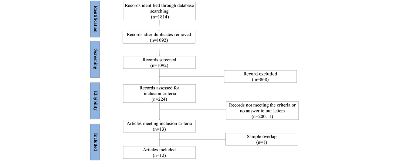
Figure 1. Meta-analysis of resting-state studies in
medication-naive patients with first-episode major depression
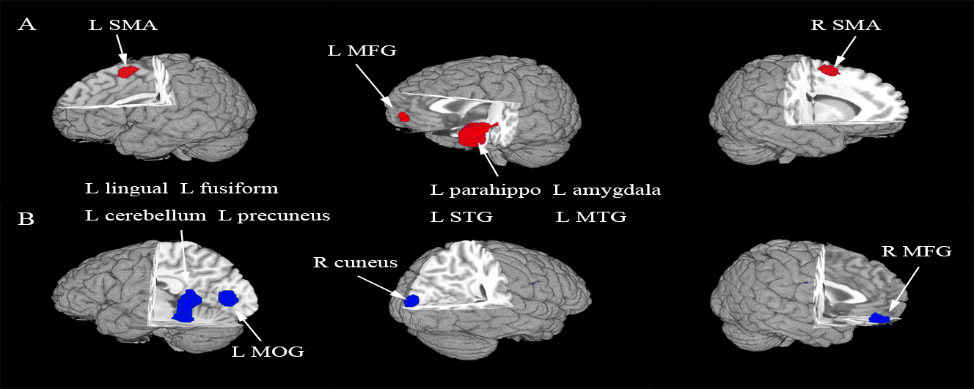
Figure 2. The areas of increased (red) and decreased (blue)
resting-state brain activity in the meta-analyses of studies in
medication-naive patients with first-episode major depression compared with
healthy controls. R, right; L, left; SMA, supplementary motor area; MFG, middle
frontal gyrus; MOG, middle occipital gyrus; STG, superior temporal gyrus; MTG,
middle temporal gyrus.