1832
Change of cortical thickness and hippocampal volume in adolescents with autism spectrum disorder1Electrical Engineering, National Sun Yat-Sen University, Kaohsiung, Taiwan, Taiwan, 2School of Medicine, National Yang-Ming University, Taipei, Taiwan, Taiwan, 3Radiology, Kaohsiung Veterans General Hospital, Kaohsiung,Taiwan, Taiwan, 4Psychiatry, Kaohsiung Medical University and Kaohsiung Medical University Hospital, Kaohsiung, Taiwan, Taiwan
Synopsis
By using a surface-based method (Freesurfer), the cortical thickness, hippocampal volume, and amygdala volume measurement were performed on adolescents with autism spectrum disorder (n=17) and age-matched typically developing controls (n=10). ASD patients showed a thicker cortex in temporal and occipital regions, a thinner cortex in frontal regions, and larger right hippocampal volume compared to the controls.
Background
Autism spectrum disorder (ASD) is a neurodevelopmental disorder that induces problem in social communication and interaction. As a powerful tool to investigate human brain, magnetic resonance imaging has been used to inspect structural changes of ASD patients. Previous longitudinal studies suggested that abnormal cortical development in ASD may follow three phases, including accelerated expansion in early childhood, accelerated thinning in late childhood and adolescence, and decelerated thinning in early adulthood. [1] Although the abnormality of cortical thickness is obviously associated with age, there is no common result of cortical thinning or thickening on specific brain area. [2] Furthermore, several imaging studies show that the amygdala and hippocampus may be associated to functional deficit in ASD. [3] [4] The goal of this study is to conduct the comparison independently and accumulate local experience by examining the anatomical change, including cortical thickness and volume of hippocampus and amygdala, of autism at age 15 to 20.Methods
Imaging data were collected at 3 T (Magnetom Skyra, Siemens, Erlangen, Germany) on seventeen adolescents diagnosed as autism according to DSM-4-TR (mean age=17.15±1.13 years old) and ten age-matched typically developing (TD) controls (mean age=16.68±1.68 years old). All subjects are male. The whole-brain T1WI was obtained by MPRAGE sequence (TE/TR = 2.07/2000 ms, TI = 900 ms, flip angle = 9°, FOV = 256 mm, voxel size = 1mm3). The volume of brain tissues and the cortical thickness are estimated by using a fully-automated surface-based method (FreeSurfer software package, version 5.3.0), which is one of the most widely used techniques at its field. In this method, the white matter-gray matter surface and the pial surface are first reconstructed. The cortical thickness at each vertex can be calculated as the distance between two mesh surfaces and mapped to the inflated cortex. The mapping of cortical thickness is spatially smoothed by a Gaussian kernel with a full width at half maximum of 20 mm. The general linear model is applied to examine the association between cortical thickness and age. Two-sampled t-test is used for group comparison between the autism and control groups. Statistically significant difference is found when the two-tailed p value is less than 0.05.Results
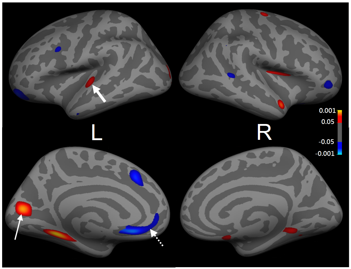
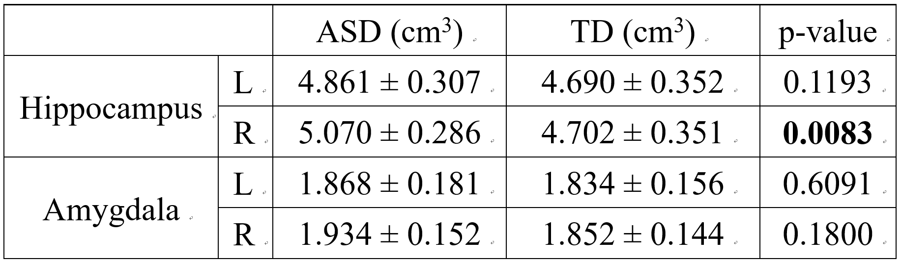
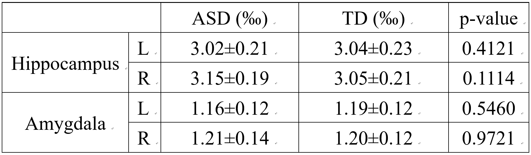
Figure 1 shows thicker (red) and thinner (blue) cortical area of ASD compared to typically developing (TD) males. Among 62 regions of Desikan-Killianny-Tourville (DKT) atlas, the autism group shows thicker cortical thickness at left pericalcarine cortex (pointed by the thin arrow) and the transverse temporal gyrus (thick arrow), and thinner cortex at rostral anterior cingulate (dotted arrow). For the comparison of volume of hippocampus and amygdala, significant difference is only found on the right hippocampus (p<0.01, see Table 1). After normalized by individual’s intracranial volume to avoid the influence of head size, the right hippocampal volume of autism is still larger than control, but the p value is no longer less than 0.05. (Table 2)Discussion
For ASD patients, this study found several thicker cortical regions on the temporal and occipital lobes, and scattered thinner cortical regions, mostly in frontal area, compared to age-matched control. In previous studies, Zielinski BA et al. also showed adolescents with ASD may have thinner cortical thickness than TD at frontal lobe. [1] Although thinner cortex was also reported on the various regions, especially in temporal and parietal lobes [2], recent analysis using multi-site online data base shows thicker cortex at left temporal and occipital regions, which reaches agreement with our results. [5] In addition, our result of larger right hippocampal volume also correspond to the study that have done on ASD children, suggesting that the difference of volume may persist until late adolescence and early adulthood. The main limitation of this study is the small sample size of participants, limiting the statistical power. Further longitudinal investigation and analysis of subgroup is required to provide more insight of cortical development of ASD.Acknowledgements
No acknowledgement found.References
[1] Brandon A. Zielinski, M.B.D.P., Jared A. Nielsen, Alyson L. Froehlich, Tracy J. Abildskov, Jeffrey S. Anderson, P. Thomas Fletcher, Kristen M. Zygmunt, Brittany G. Travers, Nicholas Lange, Andrew L. Alexander, Erin D. Bigler6, and Janet E. Lainhart, Longitudinal changes in cortical thickness in autism and typical development. BRAIN, 2014.
[2] Gregory L. Wallace, N.D., Lauren Kenworthy, Jay N. Giedd and Alex Martin, Age-related temporal and parietal cortical thinning in autism spectrum disorders. BRAIN, 2010.
[3] Naama Barnea-Goraly, T.W.F., Lucia Piacenza, Nancy J. Minshew, Matcheri S.Keshavan , Allan L. Reiss, Antonio Y. Hardan, A preliminary longitudinal volumetric MRI study of amygdala and hippocampal volumes in autism. Neuro-Psychopharmacology &Biological Psychiatry 2014: p. 124–128.
[4] Xiao-Jing Shou, X.-J.X., Xiang-Zhu Zeng, Hui-Shu Yuan, Rong Zhang, Ji-Sheng Han Ying Liu, Yan Xing, Mei-Xiang Jia, Qing-Yun Wei, Song-Ping Han, A Volumetric and Functional Connectivity MRI Study of Brain Arginine-Vasopressin Pathways in Autistic Children. Neurosci. Bull, 2017: p. 130–142.
[5] Shlomi Haar, Sigal Berman, Marlene Behrmann, Ilan Dinstein, Anatomical Abnormalities in Autism? Cerebral Cortex, 2016, Vol. 26, No. 4
Figures