1827
Alterations in amplitude of low frequency fluctuation in drug-free major depressive disorder1Huaxi Magnetic Resonance Research Centre (HMRRC), Department of Radiology, West China Hospital of Sichuan University, chengdu, China
Synopsis
The objective of this study was 1) to confirm whether the intrinsic brain activities (as evaluated by ALFF) in the anterior cingulate cortex (ACC) is associated with antidepressant treatment in a relative large sample of drug-free major depressive disorder (MDD) patients and 2) to determine whether the pretreatment ALFF activities predict the effect of the follow-up antidepressant treatment in MDD. Our findings demonstrate that intrinsic brain activities in the ACC was influenced by disease itself rather than antidepressant treatment and threw light on predictive value of the right thalamus as a marker of short term antidepressant treatment outcome in MDD.
Purpose
Many studies have examined amplitude of low-frequency fluctuation (ALFF)1 changes in the anterior cingulate cortex (ACC) of patients with major depressive disorder (MDD), however, the results were inconsistent. In addition, few studies have investigated the relationship between ALFF and outcome to antidepressant treatment. Recently, a meta-analysis of ALFF studies2 showed that increased ALFF activity in the ACC is more likely to be associated with antidepressant treatment. However, most of the original literature in this meta-analysis had a small sample size and did not find any ALFF changes in the ACC. Due to the heterogeneity of meta-analysis, the two goals of the current study are: 1) to confirm whether the ALFF activity in the ACC is associated with antidepressant treatment in a relative large sample of drug-free MDD and 2) to determine whether the pretreatment ALFF predict the effect of the follow-up antidepressant treatment in MDD.
Methods
The study was approved by the local ethical committee and written informed consent was obtained from all subjects. A total of 52 drug-free MDD patients who met DSM-IV criteria for MDD and 52 age, gender and handedness well matched healthy controls were recruited. Depressive symptom severity was assessed using the 17-item Hamilton Depression Rating Scale (HAMD). After MRI examination, antidepressant treatment trial was applied for all patients according to the clinical judgment of the treating psychiatrist as described in our previous publication3. The efficiency of medication was represented by reduction rate of HAMD score (RRS), which was calculated as [(HAMDbefore-HAMDafter6W)/HAMDbefore]. According to RRS at six weeks, patients group was further divided into two subgroups: remitted (RRS≥50%) depression (RD) group and non-remitted (RRS<50%) depression (NRD) group.
The rs-fMRI images sensitized to changes in the blood oxygen level dependent (BOLD) signal levels were obtained using EPI sequence via a 3-Telsa GE scanner. Participants were instructed to relax with their eyes closed and keep still. The ALFF maps were calculated using DPARSF software (http://www.restfmri.net) for each subject. A two-sample t-test of individual normalized ALFF maps was conducted in SPM8 (http://www.fil.ion.ucl.ac.uk/spm) between MDD and controls. The significance threshold was set at P < 0.05 (AlphaSim corrected; combined height threshold P < 0.005 and a minimum cluster size of 37 voxels). Brain areas with significant ALFF differences between groups were taken as regions of interest (ROIs) for Pearson’s correlation analyses with HAMD score or illness duration or number of episode or RRS in SPSS. In subgroup analysis, ALFF maps were compared among the RD group, NRD group and HC using analysis of variance (ANOVA). The significance threshold was set at P < 0.05 (AlphaSim corrected; combined height threshold P < 0.005 and a minimum cluster size of 37 voxels). For brain regions identified in the ANCOVA, we extracted those regions as ROIs and then performed follow-up LSD-tests in regions with overall group differences to identify pair-wise group differences using the SPSS.
Results
Demographic and clinical characteristics for all subjects are summarized in Table 1. Relative to HC, MDD patients showed increased ALFF in the ACC, cerebellum, bilateral hippocampus and decreased ALFF in the right thalamus (P < 0.05, AlphaSim corrected) (Fig 1 & table 2). In subgroup analysis, the three groups had significant differences of ALFF in the right thalamus (P < 0.05, AlphaSim corrected) (Fig 2A). Post-hoc LSD-tests showed that RD group had decreased ALFF in the right thalamus relative to both NRD group and HC (Fig 2B). Relative to controls, NRD group showed significantly decreased ALFF in the right thalamus (Fig 2B). There was a negative correlation between illness duration and ALFF values in the cerebellum in MDD (r=-0.276, p=0.047) (Fig 3).Discussion & Conclusion
The main finding of this study was that increased ALFF in the ACC showed in drug-free MDD patients who have excluded the influence of medication. Our finding was inconsistent with previous meta-analysis2 and indicated that intrinsic brain activities in the ACC was influenced by disease itself and gave an evidence that the ACC activity plays an important role in the pathophysiology of MDD. Negative correlation between illness duration and ALFF values in the cerebellum indicated that cerebellum involved in the pathologic development of MDD, which was consistent with previous meta-analysis2. In the analysis of the subgroups based on RRS showed that RD group had decreased ALFF in the right thalamus relative to NRD group, which suggested that the lower ALFF in the right thalamus may correlated with better antidepressant treatment outcome in six weeks. Our findings threw light on predictive value of the right thalamus as a marker of short term antidepressant treatment outcome in MDD. Future longitudinal study with large sample size are needed to verify this finding.Acknowledgements
No acknowledgement found.References
1. Zang, Y.F., et al. Altered baseline brain activity in children with ADHD revealed by resting-state functional MRI. Brain Dev, 2007. 29(2): p. 83-91.
2. Zhou, M., et al. Intrinsic cerebral activity at resting state in adults with major depressive disorder: A meta-analysis. Prog Neuropsychopharmacol Biol Psychiatry, 2017. 75: p. 157-164.
3. Lui, S., et al. Resting-state functional connectivity in treatment-resistant depression. Am J Psychiatry, 2011. 168(6): p. 642-8.
Figures
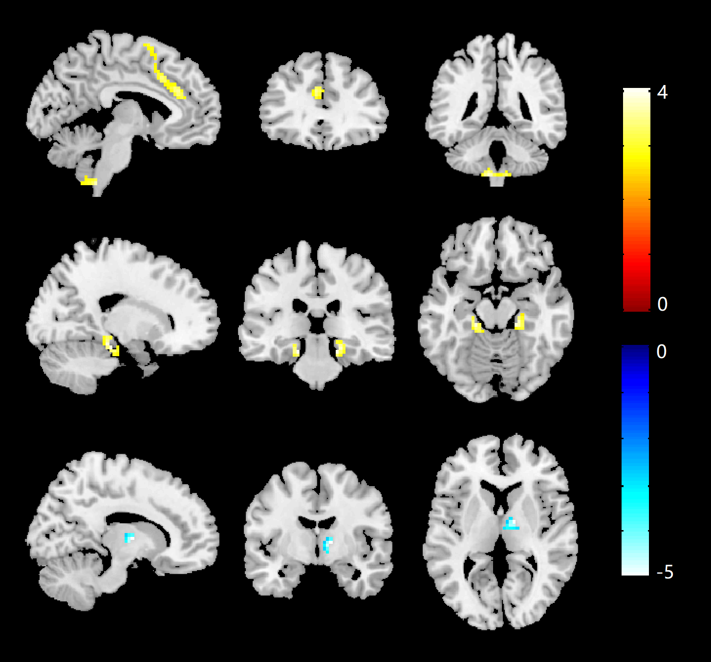
Fig 1. Alterations of ALFF in MDD patients compared with HC. ALFF increases are indicated in warm colors while ALFF decreases are indicated in cool colors. Clusters are shown after controlling for multiple comparisons with AlphaSim correction (P<0.05). The color bar represents the range of T values. Red circles, increased brain activation regions; blue squares, decreased brain activation regions.
Abbreviations: ALFF, amplitude of low frequency fluctuation; MDD, major depressive disorder; HC, healthy controls.
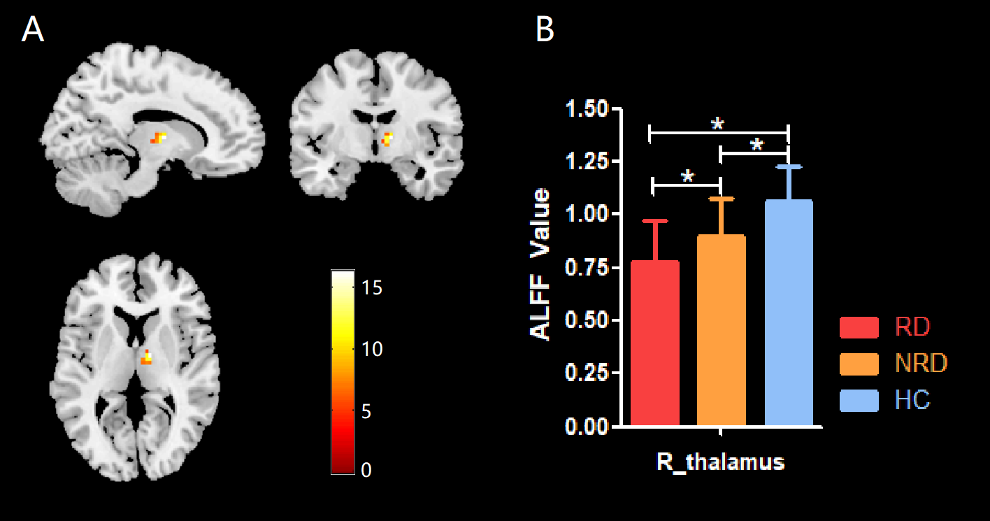
Fig 2. (A) The significant ALFF differences among RD group, NRD group and HC. Clusters are shown after controlling for multiple comparisons with AlphaSim correction (P<0.05). The color bar represents the range of F values. (B) Using these brain regions as ROIs, post hoc LSD-tests were performed to identify pair-wise group differences.
Abbreviations: ALFF, amplitude of low frequency fluctuation; HC, healthy controls; RD, remitted depression; NRD, non-remitted depression.
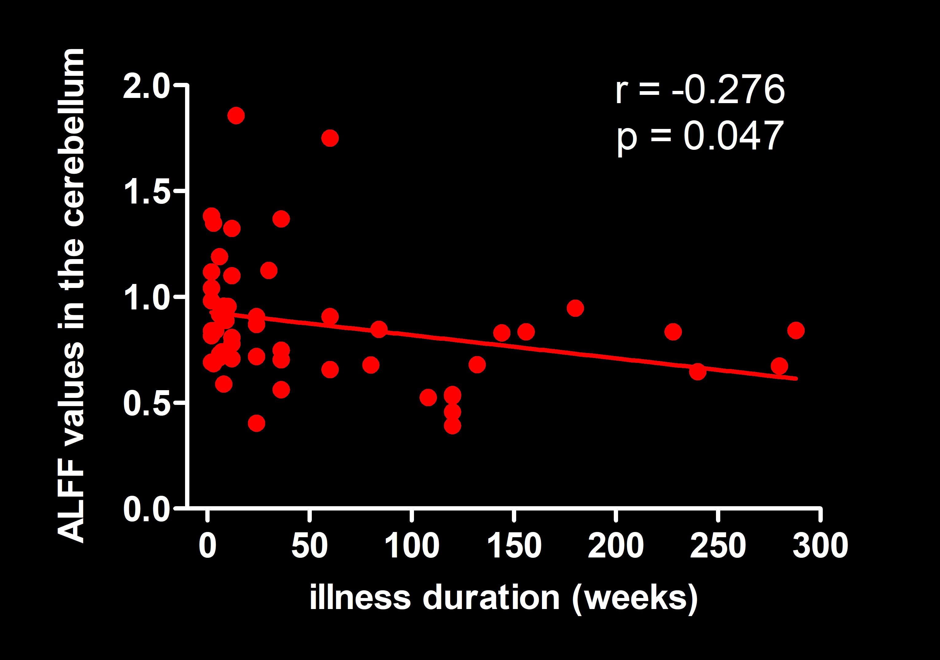
Fig 3. Pearson correlation exhibiting negative correlation between illness duration and ALFF values in the cerebellum in MDD.
Abbreviations: ALFF, amplitude of low frequency fluctuation; MDD, major depressive disorder.
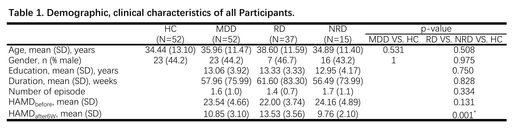
*Statistical significance between remitted depression group and non-remitted depression group was observed for the Hamilton Rating Scale for Depression score after six weeks' antidepressant treatment. (p=0.001). No other significant differences were found.
HAMDbefore indicates the Hamilton Rating Scale for Depression score before treatment.
HAMDafter6w indicates the Hamilton Rating Scale for Depression score after six weeks' antidepressant treatment.
Abbreviations: RD: remitted depression; NRD, non-remitted depression; HC, healthy controls; HAMD, Hamilton Rating Scale for Depression; SD, standard deviation.
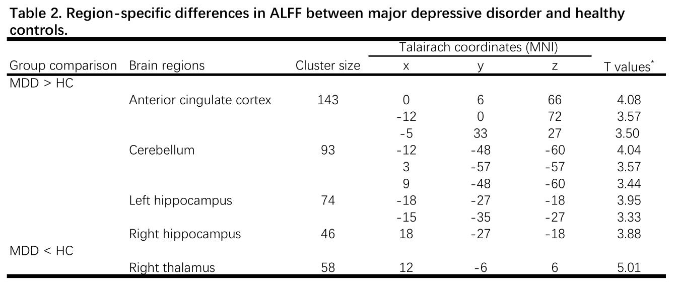
*T-statistical value of peak voxel showing ALFF differences between MDD and HC.
Data corrected for multiple comparisons with AlphaSim correction.
Abbreviations: ALFF, amplitude of low-frequency fluctuations; MDD, major depressive disorder; HC, healthy controls.