1821
The Differences of Amplitude of Low Frequency Fluctuation between Methamphetamine and Heroin use disorder: a resting-state functional magnetic resonance imaging studyYan Liu1,2, Wei Wang1, Wei Li1, Qiang Li1, Yongbin Li1, Jiajie Chen1, Jing Chen1, and Shan Dang1
1Department of Radiology, Tangdu Hospital, the Air Force Medical University, XI AN, China, 2Department of Radiology, Changqing Xinglongyuan Hospital, Affiliated Hospital of Changqing Oilfield, XI AN, China
Synopsis
These findings indicated different brain regions between MA users and heroin users in resting-state, as well as it’s function correlation with emotion.
Purpose
Methamphetamine (MA) is a powerful central nervous system stimulant of amphetamine combinations and has strong addictive properties. Chronic misuse of stimulants may damage brain cells, cause neurotoxicity and lead to long-lasting impairment in brain function. In clinic, compared with heroin, methamphetamine use disorder (MAUD) individuals show more prominent spirit dependence instead of body dependence. MAUD individuals are often associated with a series of psychiatric problems, such as schizophrenic manifestations with delusion and hallucination. It's more likely irritability, aggression, and impulsive behavior. So, we suspected MAUD individuals have special spirit impaired. At present, the brain functional magnetic resonance imaging (fMRI) study of MAUD was mainly focus on task-design pattern, correspondingly, it highlight the task-making brain areas in MA. However, it is very important to determine the tissue pattern of brain function in the baseline of the MAUD. The method of amplitude of low-frequency fluctuation(ALFF) in resting-state is an effective method. This would help to find the pathological basis of methamphetamine associated mental damage and damaged brain areas differ from heroin, and provide reliable theoretical basis of exploring effective treatment program and clear therapeutic targets.Methods
21 male MAUD individuals, 21 male heroin use disorder (HUD) individuals and 21 male demographically-matched healthy controls (HC) were recruited. The MAUD and HUD individuals were diagnosed according to the diagnostic criteria of the DSM-V. If they use other illegal drug or have psychiatric illness, they will be excluded. The method of ALFF was used to explore the causes of significant differences among three groups. All MR imaging were acquired using an eight-channel head coil on a 3.0 T GE-Signa MRI scanner (GE Healthcare, Milwaukee, U.S.A.). The differences of ALFF between these three groups were investigated by ANOVA analysis. Psychologic situation were evaluated by the Self-reporting Inventory-90(SCL-90). Pearson correlation analysis was performed to explore the relationship between the ALFF values and the psychological scores.Results
The SCL-90 scores of three groups were different in anxiety, hostility, stubborn, and core psychotic items (P<0.05, AlphaSim corrected), MA users were significantly higher than others. Two-two comparison between the three groups showed MAUD group showed higher levels of anxiety, hostility, stubborn and core psychotic compared with HC group. Additionally, those in the MAUD group had higher scores of hostility compared with HUD group. The ALFF values of multiple brain between the three groups were different and the brain regions were cerebral, thalamus, left frontal middle gyrus, right inferior temporal gyrus and bilateral posterior central (P<0.001, Alpha Sim corrected). Compared with the HUD group, the MAUD group demonstrated significantly decreased brain activity in resting-state of cerebellum and left frontal middle gyrus, but increased brain activity in thalamus, right inferior temporal gyrus and bilateral posterior central gyrus. Compared with HC group, the MAUD group demonstrated significantly decreased in cerebellum, increased in thalamus, right posterior central and right inferior temporal gyrus. The ALFF value of cerebral was negatively correlated with the anxiety subscale on the SCL-90(r=-0.446,p=0.043).Conclusions
Based on the above, we believe that methamphetamine and heroin have different effects on different brain region, it is showed that the effects on neurons in the same brain region is different. The prefrontal lobe is main brain region in addict neural circuits theory and be responsible for craving and cognitive control. The ALFF of the left frontal middle gyrus of HUD is higher than MAUD and HC. Contrary to expectation, we did not find reliable evidence between MAUD and HC groups difference in prefrontal activation during resting-state, although left frontal middle gyrus activation tended to be lower in the MAUD individuals than the healthy participants during resting-state, this difference was not significant. The thalamus is believed to act as a relay between many of subcortical areas and the cerebral cortex, playing an important role in goal-directed behaviors, reward processing, cognitive and motor functions. Temporal lobe is where the auditory speech area is located, the auditory hallucinations patient has activation of the temporal lobe and auditory cortex (frontal lobe, parietal lobe, limbic system). The neuronal basis of impulsivity and moral judgment is the junction of FPPFC and temporal lobe. The postcentral gyrus service for the mirror nervous system which is closely related to the empathy and emotional expression, therefore, impaired postcentral gyrus may be one of neural mechanism of aggressive behavior. Cerebellum is involved in addictive behavior. Our research shows differential brain region focus on the cognitive and emotional systems. Our study provide reliable theoretical basis for explore suitable, effective treatment plan and identify therapeutic target in future.Acknowledgements
No acknowledgement found.References
Figures
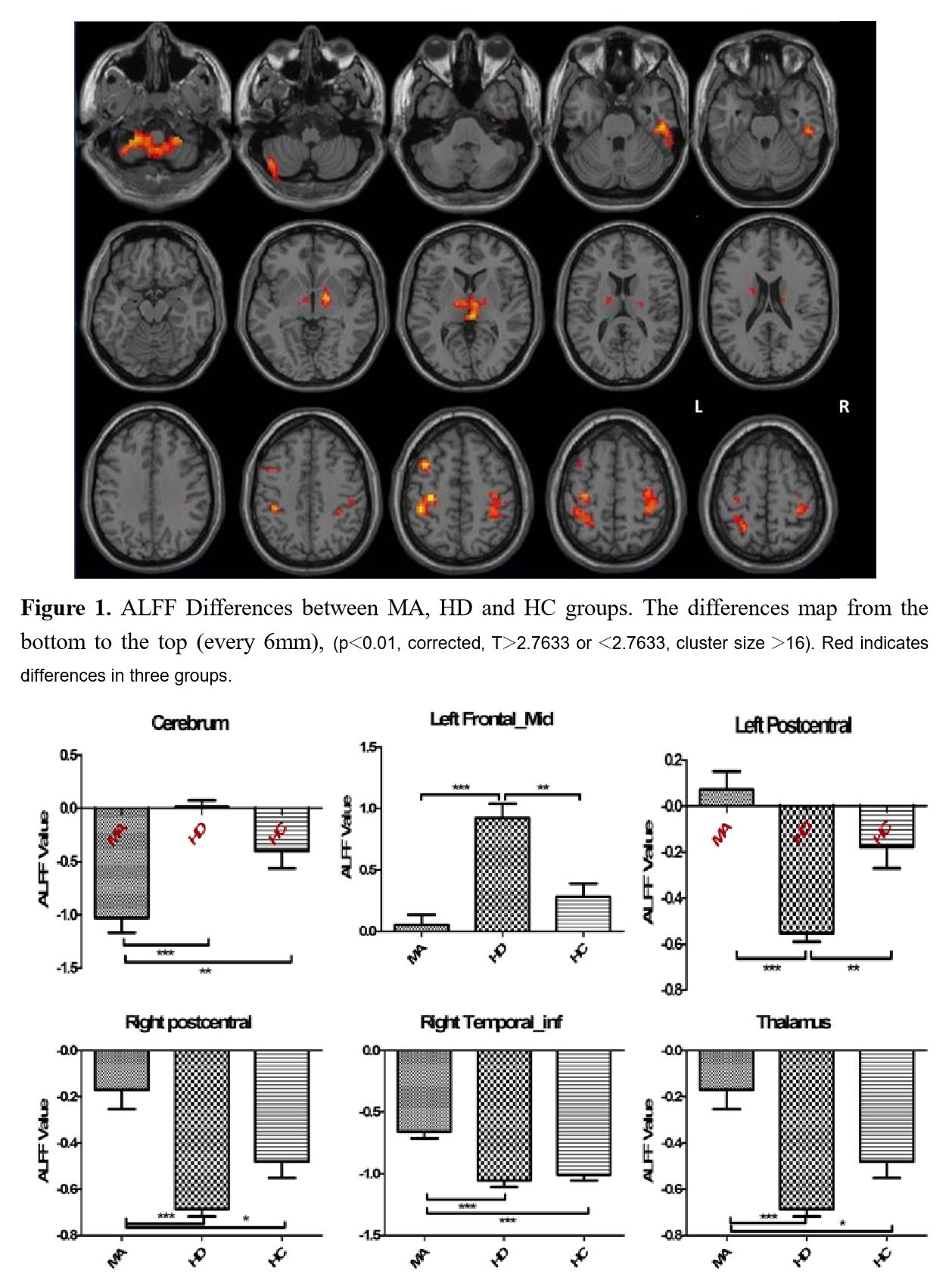
Figure 1. ALFF Differences between MA, HD and HC groups. The differences map from the bottom to the top (every 6mm), (p<0.01, corrected, T>2.7633 or <2.7633, cluster size >16). Red indicates differences in three groups.
Figure 2. The Column Diagram of ALFF Value Differences between MA, HD and HC groups. *Significant difference (P < 0.05). (*: P <0.05; **: P <0.01; ***: P <0.001)