1819
Auditory system altered in auditory verbal hallucination studied using diffusion spectrum imaging, T1-weighted image and fMRI1Department of Biological Psychiatry and Neuroscience, Dokkyo Medical University, Tochigi, Japan
Synopsis
To understand the pathology of auditory verbal hallucination (AVH), we investigated 3 MRI indices: generalized fractional anisotropy (GFA) using diffusion spectrum imaging in the auditory radiation, gray matter volume (GMV) using T1-weighted images in Heschl’s gyrus (i.e., auditory cortex) and BOLD contrast estimates using task-fMRI in the auditory cortex. The BOLD relative to the GFA was significantly greater in controls than in patients with schizophrenia who had AVH. The GMV relative to the GFA also tended to show greater values in controls than in patients. An unregulated auditory sensation attributed to a dysfunction in the cortex might eventually encompass AVH.
INTRODUCTION
Auditory verbal hallucination (AVH) is a devastating symptom of schizophrenia. We investigated alterations in the auditory system in AVH using 3 types of MRI technologies: diffusion spectrum imaging (DSI) for the auditory radiation (AR), T1-weighted image for gray matter volumes (GMV) in Heschl’s gyrus and fMRI for the estimations of functional activity in the auditory cortex (i.e., Heschl’s gyrus). Two hypotheses, which were mutually exclusive, were possible. One was a “fiber-first” hypothesis, supposing that an impairment in the AR would cause a reduction in the GMV in Heschl’s gyrus, and then result in aberrant activity in the auditory cortex. Previous studies appeared to support this hypothesis1-3. However, an opposite hypothesis was also possible, supposing that the dysfunction in the auditory cortex with a reduction of the GMV and the activity would cause a failure to control the AR, inducing AVH (“cortex-first” hypothesis).
METHODS
[Subjects]
Four patients with schizophrenia who had AVH (age 25-54) (diagnosed with DSM-V) and age- and gender-matched 8 controls (age 25-51) participated in this study. The severity of patients’ AVH was measured using PSYRATS-J. The total scores ranged from 28 to 32 (out of 44 points).
[Imaging and Analysis]
We used 3-T MRI with a 32-ch head coil (GE Healthcare).
DSI was conducted using the following parameters: 56 axial slices interleaved, TR 8000, TE minimum, slice thickness 2.5 mm, FOV 200, 80 x 80, max b-value 4000 sec/mm and 102 directions (half sphere). We computed generalized fractional anisotropy (GFA) in the whole brain. An LDDMM algorithm generated a group template to transform the individual brains to the template brain and to extract fiber tracts4-5. In the auditory radiation, 25 out of 100 steps of the GFA adjacent to Heschl’s gyrus were averaged and used as the index of diffusion anisotropy.
T1-weighted images were taken by parameters of: 170 sagittal slices, TR 8.2, TE 3.2, FA 12, TI 450, slice thickness 1 mm, FOV 256 and 256 x 256. Images underwent segmentation into gray matter and other structures using a DARTEL algorithm in SPM12. Voxel values of normalized, modulated and smoothed gray matter images within Heschl’s gyrus were averaged to use as the index of GMV.
Two task-fMRI runs in a block design were conducted by the following parameters: 45 axial-coronal slices interleaved, TR 2000, TE 22, FA 90, slice thickness 3 mm, FOV 192, 64 x 64 and 136 volumes. Eight task and 9 fixation blocks occurred alternately (total 272 sec). One run conducted a narration listening task, whereas the other run conducted a button press task to auditory cues. We included both tasks into a design matrix using SPM12. Averages of contrast estimates (task greater than fixation) extracted from Heschl’s gyri were used as the index of activity (BOLD).
[Statistics]
We performed a 2-way ANOVA for each index (between factor: schizophrenia/controls, within factor: left/right). We employed generalized omega square to estimate the effect size (ES).
RESULTS
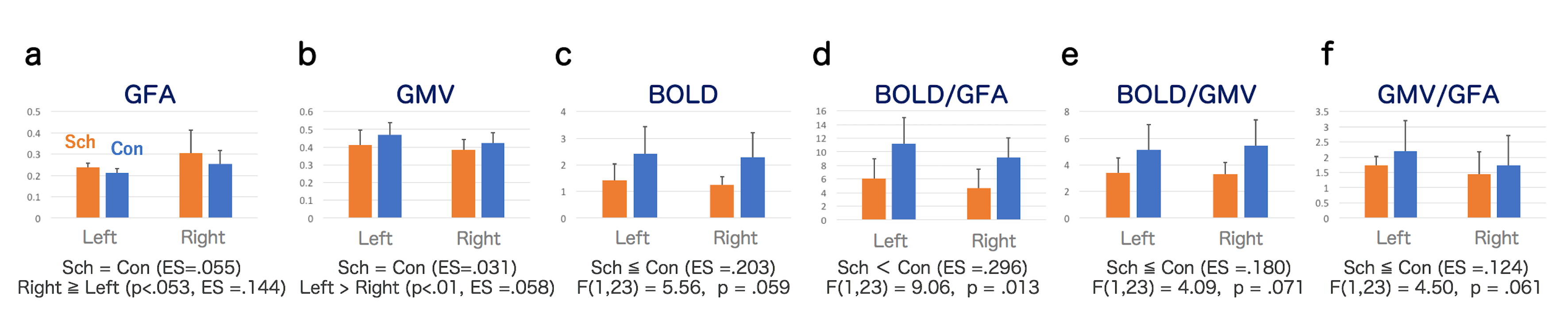
We found an accordance with the “cortex-first” hypothesis in general (Fig.1). The activity in Heschl’s gyrus tended to be extensive in controls compared to patients (F(1,23)=5.56, p=.059, ES=.203) (Fig.1c). The GMV (ES=.031) (Fig.1b) and the GFA (ES=.055) (Fig.1a) demonstrated no significant differences between patients and controls. To estimate the dominancy between fiber and cortex, we computed ratios of “BOLD divided by GFA”, “BOLD divided by GMV” and “GMV divided by GFA”. Among these, the BOLD/GFA was significantly greater in control than in patients (F=9.06, p=.013, ES=.296) (Fig.1d). The BOLD/GMV (ES=.180) (Fig.1e) and GMV/GFA (ES=.124) (Fig.1f) indicated tendencies of controls greater than patients (p<.10).DISCUSSION
The decrease of the BOLD in schizophrenia (Fig.1c) suggested a dysfunction in the auditory cortex that might affect the AVH. The GMV also suggested a reduction in schizophrenia2 (Fig.1b), albeit no statistical significance. In contrast, patients showed slightly greater GFA in the AR than controls (Fig.1a), which caused smaller BOLD/GFA (Fig.1d) and GMV/GFA (Fig.1f) in patients than in controls. These indicated a decrease of BOLD relative to GFA as well as GMV, and a decrease of GMV relative to GFA, in patients as compared to controls. Thus, the results suggested an overall dominancy of the cortex (Heschl’s gyrus, i.e., auditory cortex) over the fiber tract (AR) in healthy controls. The dysfunction in the cortex would make the auditory sensation in the AR unregulated. The out-of-control sensation might eventually encompass an AVH in schizophrenia.CONCLUSIONS
A dysfunction in Heschl’s gyrus, which exhibits a reduction of the gray matter volume and a decrease of the activity, would induce an out-of-control condition in the auditory system and an increase in the diffusion anisotropy of the auditory radiation. The alteration would eventually elicit auditory verbal hallucination in schizophrenia.Acknowledgements
- JSPS KAKENHI Grant Number 15K15428 (Japan)
- Dept. Psychiatry (patients) and Dept. Radiology (MRI), Hamamatsu University School of Medicine
- National Center of Neurology and Psychiatry, National Institute of Mental Health (Japan) (PSYRATS-J)
- Institute of Medical Device and Imaging, National Taiwan University (DSI Toolkit)
- GE Japan (introduction of DSI)
References
- Woo PY, Leung LN, Cheng ST, et al. Monoaural musical hallucinations caused by a thalamocortical auditory radiation infarct: a case report. J Med Case Rep. 2014;8:400.
- Modinos G, Costafreda SG, van Tol MJ, et al. Neuroanatomy of auditory verbal hallucinations in schizophrenia: a quantitative meta-analysis of voxel-based morphometry studies. Cortex. 2013;49(4):1046-1055.
- Jardri R, Pouchet A, Pins D, et al. Cortical activations during auditory verbal hallucinations in schizophrenia: a coordinate-based meta-analysis. Am J Psychiatry. 2011;168(1):73-81.
- Hsu YC, Hsu CH, Tseng WY. A large deformation diffeomorphic metric mapping solution for diffusion spectrum imaging datasets. Neuroimage. 2012;63(2):818-834.
- Chen YJ, Lo YC, Hsu YC, et al. Automatic whole brain tract-based analysis using predefined tracts in a diffusion spectrum imaging template and an accurate registration strategy. Hum Brain Mapp. 2015;36(9):3441-3458.
Figures