1814
Principal Component Analysis of Schizophrenia Reveals Link Between Auditory Hallucination Severity and Fractional Anisotropy in the Corpus Callosum1Psychiatry, University of British Columbia, Vancouver, BC, Canada, 2Pediatrics, University of British Columbia, Vancouver, BC, Canada
Synopsis
A PCA analysis of fractional anistropy (FA) was conducted from a sample of schizophrenia patients (n=42) and healthy controls (n=40) resulted in three major components: “corpus callosum”, “internal capsule/temporal/brainstem”, and “corona radiata”. Average component scores did not differ as a function of group, but a correlation of PSYRATS scores and principal components revealed the frequency, amount of distress associated with voices, and disruption associated with voices correlated significantly with the corpus callosum component. Our findings suggest that reduced interhemispheric connectivity of the prefrontal cortex is related to hallucination severity in schizophrenia, perhaps mediated through top-down processes such as source monitoring.
Introduction
Schizophrenia is considered a disorder of aberrant connectivity of large-scale networks. The study of white matter abnormalities using diffusion tensor imaging (DTI) may provide a better understanding of the disconnectivity associated with the disorder. Previous research has identified WM abnormalities in the corpus callosum and other white matter tracts in schizophrenia [1-4], but the precise nature and clinical correlates of these abnormalities have been mixed [1]. The common use of region-of-interest-based approaches has limited investigators’ ability to detect white matter abnormalities using whole-brain, data-driven research. In the current study, we applied principal component analysis (PCA) to DTI data from 42 patients with schizophrenia and 40 healthy controls to examine the relation of diagnosis and symptom severity with data-driven patterns of structural integrity. Post hoc analyses indicated that FA values in the genu of the corpus callosum correlated negatively with hallucination severity.Methods
Subjects: 42 patients with schizophrenia and 40 healthy controls were enrolled and scanned for this study. Schizophrenia symptom ratings were determined using the Signs and Symptoms of Psychotic Illness (SSPI) and the Psychotic Symptom Rating Scales (PSYRATS) tests. Note that three patients did not have PSYRATS ratings completed.
Data Acquisition: diffusion scans were acquired as follows: TR=7,088ms, TE=60ms, flip angle=90°, acquisition matrix=100x100, 70 slices, FOV=224x224x154mm3, voxel size= 2.4x2.4x2.2mm3, 60 unique diffusion directions optimized on the whole sphere, b-factor=700s/mm2, one b0 volume acquired.
Image Analysis: Diffusion data were eddy current and motion corrected using FSL's `eddy`, and converted to a diffusion tensor model using FSL's `dtifit`. Nonlinear registration to standard space, mean FA calculation, and FA skeletons were created using FSL's `TBSS`. PCA analyses of skeletonized FA values was then performed.
Statistics: Independent t-tests were conducted on participants’ component scores. Correlations were run between schizophrenic patients’ component scores and symptom ratings (SSPI and PSYRATS).
Results
Average FA value across voxels did not differ between groups.
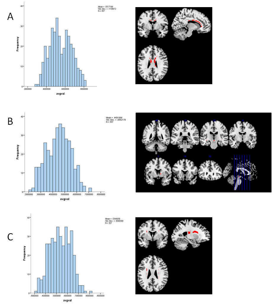
Unconstrained PCA of FA skeleton revealed 3 components (Figure 1). Component 1, “corpus callosum” was dominated by the genu and body of the corpus callosum and accounted for 11% of the FA variance. Component 2, “internal capsule/temporal/brainstem” was dominated by voxels within white matter tracts including the anterior limb of the internal capsule and the rentrolenticular part of the internal capsule, and accounted for 8% of the FA variance. Component 3, “corona radiata” was dominated by voxels in the anterior and superior corona radiata, and accounted for 5.5% of the FA variance.
Average component scores did not differ as a function of diagnosis (p’s > .43), indicating that the relative contribution of each FA pattern to overall FA variance was similar for patients and controls.
Setting a conservative alpha of .01 to control for multiple comparisons, no significant correlations emerged between SSPI symptom severity and component scores for the corpus callosum, internal capsule, or corona radiata component.
Several correlations between PSYRATS and component scores emerged, however. In patients with schizophrenia, the frequency (r=0.41), amount of distress associated with voices (r=0.47), and disruption associated with voices (r=0.41) correlated significantly with the corpus callosum component.
Discussion
Specific aspects of the phenomenology of auditory hallucinations predicted higher component scores for the corpus callosum FA component. Note that the loadings for this component were negative. This suggests that as the strength of the component increases (i.e., structural integrity in the corpus callosum decreases) there is an increase in the frequency, amount of distress associated with voices, and disruption associated with auditory hallucinations. Follow-up exploratory correlations found that indeed, average FA values in the corpus callosum-- particularly the anterior corpus callosum-- correlated negatively with hallucination severity and qualitative aspects of hallucinations (e.g., frequency, distress).
Probabilistic tractography in the HARDI template of the IIT Human Brain Atlas found that the white matter voxels dominating the corpus callosum component projected primarily to bilateral prefrontal regions. Together, these findings suggest that reduced interhemispheric connectivity of the prefrontal cortex is related to hallucination severity in schizophrenia, perhaps mediated through top-down processes such as source monitoring.
Acknowledgements
No acknowledgement found.References
1. Ellison-Wright, I., & Bullmore, E. (2009). Meta-analysis of diffusion tensor imaging studies in schizophrenia. Schizophrenia research, 108(1), 3-10.
2. Patel, S., Mahon, K., Wellington, R., Zhang, J., Chaplin, W., & Szeszko, P. R. (2011). A meta-analysis of diffusion tensor imaging studies of the corpus callosum in schizophrenia. Schizophrenia research, 129(2), 149-155.
3. Bora, E., Fornito, A., Radua, J., Walterfang, M., Seal, M., Wood, S. J., ... & Pantelis, C. (2011). Neuroanatomical abnormalities in schizophrenia: a multimodal voxelwise meta-analysis and meta-regression analysis. Schizophrenia research, 127(1), 46-57.
4. Kubicki, M., McCarley, R., Westin, C. F., Park, H. J., Maier, S., Kikinis, R., ... & Shenton, M. E. (2007). A review of diffusion tensor imaging studies in schizophrenia. Journal of psychiatric research, 41(1), 15-30.
Figures