1807
Hippocampus and parietal lobe glutamate changes as a function of age in schizophreniaFrank E. Gaston1, S. Andrea Wijtenburg1, Stephanie A. Korenic1, Hongji Chen1, and Laura M. Rowland1
1Psychiatry, University of Maryland School of Medicine, Baltimore, MD, United States
Synopsis
MRS was used to examine the aging effects of glutamate in participants with schizophrenia versus healthy controls. The parietal lobe and hippocampus, regions associated with general aging and the pathophysiology of schizophrenia, were assessed. Results revealed that hippocampal glutamate was lower in older adults with schizophrenia versus older controls. In contrast, parietal glutamate was lower in schizophrenia versus controls, irrespective of age group. These results suggest that the hippocampus may be particularly vulnerable to aging in schizophrenia. Interventions that halt hippocampal glutamate decline may be beneficial for patients with schizophrenia.
Target Audience
Researchers interested in studying schizophrenia using MRSIntroduction
Glutamate, the primary excitatory neurotransmitter in the human brain, may decline with age at a greater rate in schizophrenia compared to the general population as suggested by meta-analysis1. A recent magnetic resonance spectroscopy (MRS) study examining the anterior cingulate showed that glutamate declined with age at similar rates in both schizophrenia and healthy controls2. However, it is unclear whether this pattern is isolated to a specific brain region or widespread to other brain regions. Here, we used MRS to investigate glutamate levels in the parietal lobe and hippocampus, regions implicated in the pathophysiology of schizophrenia and general aging.Methods
All data were acquired on a Siemens TIM Trio 3T MR system with a 32-channel head coil. The study was approved by the UMB IRB. All participants provided written informed consent. Eighty-six participants completed the study. Participants with schizophrenia were evaluated for psychopathology, and all participants completed cognitive testing with a focus on relational memory, which is hippocampal dependent. MPRAGE images were acquired in order to prescribe the MRS voxels in the hippomcampus (VOI~4.5cm3) and parietal lobe (VOI~6cm3) (Figures 1 & 2). Data were acquired using PRESS: TR/TE=2000/30ms, NEX=256/hippocampus and 128/parietal, spectral width=2.5kHz, and 2048 complex points. A water reference (NEX=16) was acquired as well for phase and eddy current correction. Glutamate was quantified using LCModel3, and Cramer Rao Lower Bounds (CRLB) standard deviation (SD) thresholds were ≤ 20% for glutamate. All data were corrected for the proportion of proportion of gray matter, white matter, and CSF within the spectroscopic voxel using Matlab code based on the work of Gasparovic et al4. Metabolite levels were reported in institutional units. Two-way ANOVAs with diagnosis and age as main effects were computed. Correlation analyses were conducted between glutamate and relational memory measures.Results
Representative spectra from each region as well as the corresponding LCModel fit and residual are shown in Figures 3 & 4. Hippocampal glutamate was lower in schizophrenia versus healthy controls (p=0.041). More specifically, glutamate was significantly lower in older adults with schizophrenia compared to healthy controls (p=0.005), and this was not observed when comparing young adults with schizophrenia and healthy controls (p=0.844). In the parietal lobe, glutamate was lower in older versus younger groups (p<0.001). . Parietal glutamate was lower in participants with schizophrenia compared to controls (p=0.042) irrespective of age group. Better relational memory performance was related to higher levels of parietal glutamate (p<0.01). There were no significant relationships between glutamate levels in either region and severity of psychiatric symptoms.Discussion
These results, in conjunction with previous work2, suggest that anterior cingulate and parietal cortical regions do not show accelerated aging in schizophrenia but do show reduced glutamate levels in the illness. In contrast, the hippocampus may be particularly vulnerable to aging in schizophrenia since lower hippocampal glutamate levels were observed only in older participants with schizophrenia. Interventions targeted at increasing levels in the anterior cingulate and parietal cortex and halting the decline in the hippocampus may be beneficial for patients with schizophrenia.Acknowledgements
This study was supported by the National Institute Health: R01MH094520.References
1Marsman A et al. Glutamate in schizophrenia: a focused review and meta-analysis of 1H-MRS studies. Schizophr Bull 2013. 39:120-9. 2Wijtenburg SA et al. Altered glutamate and regional cerebral blood flow levels in schizophrenia: a 1H-MRS and pCASL study. Neuropsychopharmacology. 2017. 42:562-71. 3Provencher SW. Estimation of metabolite concentrations from localized in vivo proton NMR spectra. Magn Reson Med 1993;30(6):672-679. 4Gasparovic C et al. Use of tissue water as a concentration reference for proton spectroscopic imaging. Magn Reson Med 2006; 55(6): 1219-1226.Figures
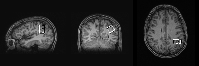
Figure
1 – Parietal Lobe
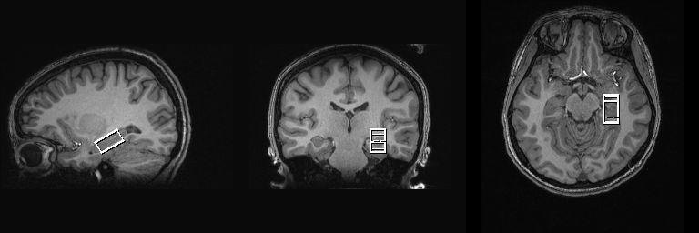
Figure
2 – Hippocampus
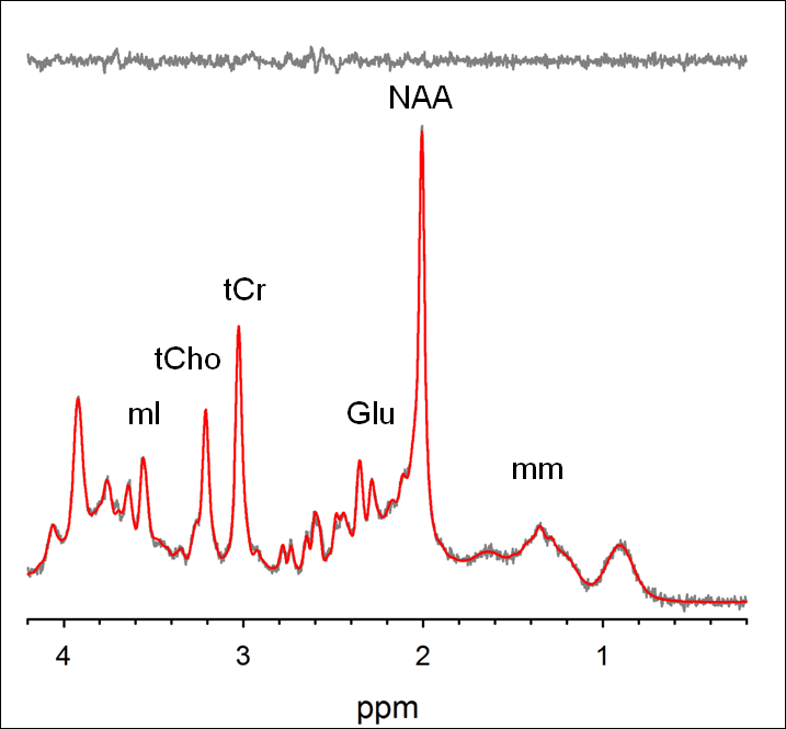
Figure
3 – Parietal Lobe
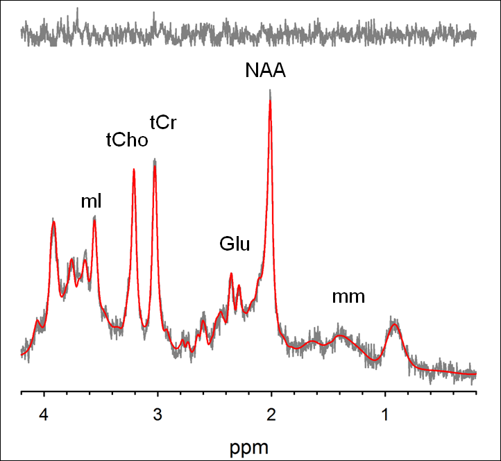
Figure
4 – hippocampus