1805
Acutely treated antipsychotics haloperidol enhances BOLD responses to the somatosensory stimulation in anesthetized rats.1Center for Neuroscience Imaging Research (CNIR), Institute for Basic Science (IBS), Suwon, Republic of Korea, 2Department of Biomedical Engineering, Sungkyunkwan University, Suwon, Republic of Korea
Synopsis
The use of BOLD fMRI is rapidly increasing for probing the effects of antipsychotics in schizophrenia. Since fMRI BOLD is an indirect measurement of neural activities, it is critical to examine the effect of antipsychotics on neurovascular coupling to prevent misinterpretation of MR data. Acutely treated haloperidol (0.2mg/kg, i.v.) increased BOLD fMRI to the somatosensory stimulation in the 1.5% isoflurane-anesthetized rats (n=5). In parallel with the BOLD results, evoked CBF and LFP by somatosensory stimuli were increased after haloperidol administration (n=8). Our results indicate that acutely treated haloperidol could influence somatosensory responses and the increased BOLD signal is coupled with enhanced neural activities.
Introduction
Dopamine is involved in multiple brain functions and malfunction of dopamine system strongly linked with several brain diseases such as schizophrenia. However, the effects of the widely used antipsychotics and a typical D2 antagonist haloperidol on sensory processing were not apparent. A haloperidol did not influence evoked CBF by sensory stimulation.1 However, differently modulated vascular activities were also reported after haloperidol treatment.2 Furthermore, specific blocking of dopamine receptor enhanced neural responses and BOLD signal to the sensory stimulation.3, 4 This confusion can be attributed to the fact that dopamine affects not only nervous system but also vascular system.5 To address this question, we executed BOLD fMRI and simultaneous recording of CBF and LFP to the sensory stimulation before and after haloperidol treatment (0.2mg/kg i.v.) in anesthetized rats.Methods
Animal preparation. All experimental procedures were approved by the Sungkyunkwan University Institutional Animal Care and Use Committee. SD rats (n=5 for fMRI, and n=8 for CBF and LFP) were anesthetized with isoflurane (~1.5%) and artificially ventilated. To monitor blood pressure continuously and inject a drug, femoral cannulation was executed as a surgical preparation. Two needle electrodes were carefully inserted into between digit 2 and 4 in forepaw to deliver electrical forepaw sensory stimuli (10Hz, 1mA).
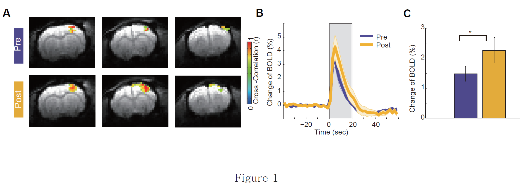
BOLD fMRI experiment. All fMRI data were acquired on a 9.4T Bruker scanner using GE-EPI sequence (TR/TE=2000/15ms, image matrix=64x32, 9 slices with 1mm slice thickness). A 72mm volume coil was used for RF transmission and 20 mm surface coil for RF receiving (Bruker BiopSpec system) was positioned on top of the rat’s head for imaging. The seven times of sensory stimulation were executed before and after haloperidol injection (Sigma, 0.2mg/kg i.v.). Twenty seconds of electrical forepaw stimulation (10Hz, 0.3ms pulse, 1mA) was delivered in each trial.
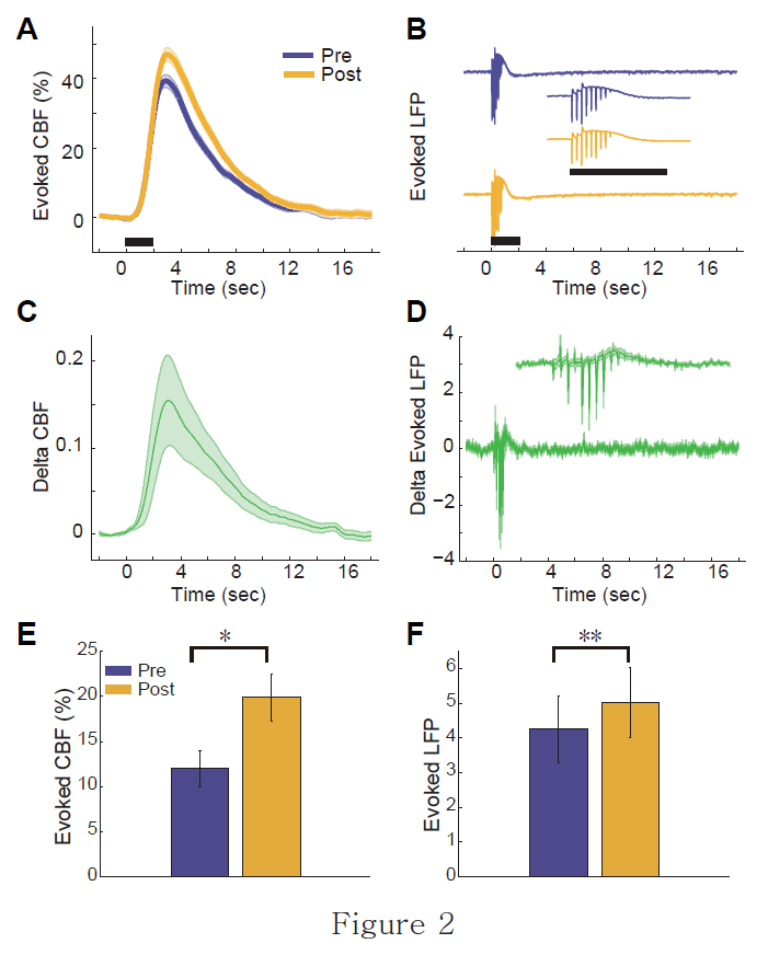
CBF and LFP experiment. Skull was drilled in the contralateral primary somatosensory cortex area (S1). A needle types laser Doppler flowmetry (LDF) probe (0.45mm diameter, 0.15mm separation, Probe 411) was carefully placed on the surface of the somatosensory cortex. A tungsten microelectrode (0.3~1 MΩ, FHC, Inc., Bowdoinham, ME) was inserted into 0.6~0.8mm below the activation cortical surface near the LDF probe. A 30 and 60 times of sensory stimulation were executed during before and after haloperidol injection (0.2mg/kg i.v.). A two seconds of electrical stimulation (10Hz, 1ms pulse, 1mA) was delivered in each trial.
Results
The acute haloperidol treatment increased evoked BOLD, CBF and LFP signals for the somatosensory stimulation. The normalized BOLD signal in an ROI to the forepaw somatosensory stimuli increased from 1.33 ± 0.23 to 2.04 ± 0.39 % after acute haloperidol treatment (n=5, paired t-test t4=-3.32, p < 0.05). Consistent with the BOLD results, both of evoked CBF and LFP were increased after acute haloperidol injection. Average amplitude and peak of evoked CBF was increased from 11.9 ± 2.0 to 19.8± 2.5 % (n=8, paired t-test, t7=-3.29, p < 0.05) and 29.4 ± 4.9 to 45.6 ± 6.1 % (n=8, paired t-test, t7=-3.17, p < 0.05). Furthermore, The evoked ∑LFP also increased from 4.25 ± 0.97 to 5.01 ± 1.01 mV after acute haloperidol (n=8, paired t-test, t7=-3.51, p<0.01). This increment of neural activities was by the first half ten pulses (n=8, paired t-test, t7=-3.32, p<0.05) but by the last half ten pulses (n=8, paired t-test, t7=-0.89, p=0.40).Conclusions
Our results indicate that acutely treated haloperidol could influence somatosensory responses and the increased BOLD signal is coupled with enhanced neural activities. This study sheds light on the underlying mechanism responsible for the effects of the antipsychotics drug on BOLD signals. Also, preclinical investigation for the chronic effect of antipsychotics on the neurovascular coupling will be needed to understand the effect of antipsychotics on BOLD in schizophrenia patients.Acknowledgements
This work was supported by IBS-R015-D1References
1. Esaki T, Itoh Y, Shimoji K, et al. Effects of dopamine receptor blockade on cerebral blood flow response to somatosensory stimulation in the unanesthetized rat. The Journal of pharmacology and experimental therapeutics. 2002;303(2):497-502.
2. Brassen S, Tost H, Hoehn F, et al. Haloperidol challenge in healthy male humans: a functional magnetic resonance imaging study. Neuroscience letters. 2003;340(3):193-6.
3. Hosp JA, Hertler B, Atiemo CO, et al. Dopaminergic modulation of receptive fields in rat sensorimotor cortex. Neuroimage. 2011;54(1):154-60.
4. Ferenczi EA, Zalocusky KA, Liston C, et al. Prefrontal cortical regulation of brainwide circuit dynamics and reward-related behavior. Science. 2016;351(6268):aac9698.
5. Krimer LS, Muly EC, Williams GV, et al. Dopaminergic regulation of cerebral cortical microcirculation. Nature neuroscience. 1998;1(4):286-9.
Figures