1794
Alterations in brain connectivity during olfaction in impulsive children1Faculty of Mathematical and Physical Sciences, BUAP, Puebla, Mexico, 2Imaging Department, Hospital Infantil de México, Federico Gómez, Mexico City, Mexico, 3UAM Iztapalapa, Mexico City, Mexico, 4Neurology Department, Hospital Infantil de México, Federico Gómez, Mexico City, Mexico
Synopsis
Impulsivity is a multi-dimensional construct of behaviors. Here we compared two cohorts of impulsive and control children. Both groups underwent a functional magnetic resonance imaging experiment which food related odor cues. Activations were larger for the impulsive group in: temporal lobe, cerebellum, supplementary motor area, frontal cortex, medial cingulate cortex, insula, precuneus, precentral, para-hippocampal & clacarine. Connectivity results showed that emotional reward based on the smell and processed in temporal lobes was the main cue driving impulsive children. This was followed by a focused attention and sensations of comfort and happiness modulated by precuneus and cingulum.
Introduction
Impulsivity is a multi-dimensional construct of behaviors which include: ineffective impulse control, premature decision making and inability to delay gratification (1). An impulsive child has increased possibilities of being a gambler, abusing illegal substances like alcohol or indulging in binge eating. It is known that odor/smell is one of the principal cues for the appearance and control of appetite as well as substance abuse (2). It is because of this that understanding the neuropsychological correlates of impulsivity through a smell stimulus, is of interest (3).Methods
Here we compared two cohorts (n=18 each) of impulsive and control children with ages between 10 and 16 years (age, sex and BMI matched). Impulsivity was confirmed through Go/No-Go Task tests. Both groups underwent a functional magnetic resonance imaging (fMRI) experiment in which odor cues were presented to them (Chocolate, Clove and Lavender). Chocolate presented a high calorie content and high reward meal, Clove represented healthy foods and Lavender a non-related to food smell. Differences in activated regions (BOLD experiment) as well as inter regional correlations (Connectivity Experiment) presented the different strategies that the two groups used to process odor related stimuli. Paradigm: Volunteers for this study visited the research center in two occasions. During their first visit, anthropometric measurements, Go/No-Go task psychological testing and clinical evaluations were performed. If volunteers fulfilled all inclusion criteria for this study, they then visited the MR unit on a second day. All imaging was performed between 7 and 10 am in the mourning and patients were asked to have had a light breakfast (they were not fasted). Once in the MR, volunteers underwent: MR sequence preparations, fMRI studies (BOLD) and anatomical imaging. The whole imaging process took a total time of 20 minutes approximately. MRI: Imaging was performed in a 1.5T Philips-Intera Achevia scanner with a NOVA gradient system set. Head birdcage coil with SENSE technology and 8 channels were used for fast imaging. BOLD imaging consisted of the acquisition of 278 brain volumes with a TR=3000 ms. An T2* weighted gradient echo sequence was used with TE=30 ms, flip angle=80°. 35 consecutive axial slices (without gaps) covering the whole infant brain from frontal lobe to cerebellum. Image resolution of 3.05 mm in plane and 4.5 mm thick were obtained with 80*80*35 matrixes. Anatomical images matched the position and volume studied by those of BOLD imaging and were obtained with a fast T1 weighted gradient echo sequence (TR=307.81 ms, TE =2.48 ms, flip angle=80°). Resolution for these images was 0.38*0.38*4.5 mm with a 640*640*35 matrix. Four whole brain acquisitions were obtained and averaged (NE=4). Image Analysis: MR images were analyzed using Matlab. Two kinds of analysis were performed. First, for the BOLD study of brain areas activated using SPM12. For connectivity analysis, Conn software was used.Results
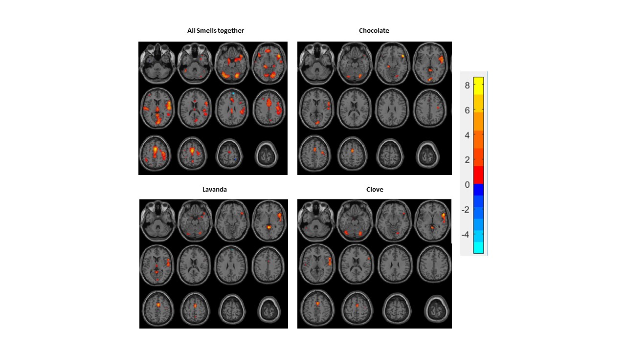
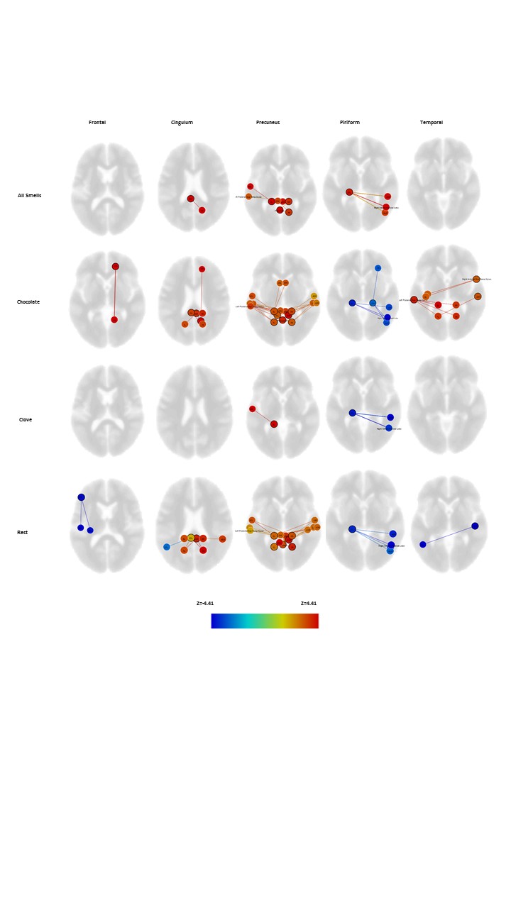
This study showed that the impulsive group had larger BOLD activations (see Figure 1) than the control in: Temporal lobe, cerebellum, supplementary motor area, frontal cortex, medial cingulate cortex, insula, precuneus, precentral, para-hippocampal & clacarine. Connectivity of these regions was then assessed and showed large amount of differential connections for the chocolate smell in: Precuneus, temporal and frontal lobes, cingulum, piriform and somatosensory cortex. Connectivity results are presented divided into these structures for clarity (Figure 2). It can be seen for the chocolate smell that impulsive volunteers presented reduced connectivity from piriform cortex to frontal and limbic structures even if their BOLD activation was larger. This reduced logical inputs from frontal lobe to the network. Precuneus (self-awareness, reward, happiness ad comfort) played a pivotal role with connections to temporal lobes (emotional memory and smell processing) implying that reward was anticipated by the chocolate smell for impulsive children but not in the control group. Cingulum together with precuneus forms part of default network and their function is anticorrelated with it. So large activations and connectivity implied these regions helped impulsive children focus on the chocolate smell (not the other odor cues).Discussion & Conclusions:
In the end we propose that impulsive subjects
have a problem in smell regulation at a neural circuit level. Impulsive
children don´t make rational decisions (no connectivity to frontal cortex) or
integrate/decipher information from other senses (touch, vision, auditory, etc.
in parietal cortex) or smell (piriform cortex) properly. They work based on emotional
memories of reward stored in temporal lobes, and auto-perceived sensations of
happiness comfort and wellbeing processed in precuneus. Finally, precuneus
together with the cingulum; keep impulsive subjects focused on the smell.Acknowledgements
NoneReferences
1. Kalenscher, T., T. Ohmann, and O. Gunturkun, The neuroscience of impulsive and self-controlled decisions. Int J Psychophysiol, 2006. 62(2): p. 203-11.
2. Yeomans, M.R., Olfactory influences on appetite and satiety in humans. Physiol Behav, 2006. 89(1): p. 10-4.
3. Heinz, A., et al., Alcohol Craving and Relapse Prediction: Imaging Studies, in Advances in the Neuroscience of Addiction, C.M. Kuhn and G.F. Koob, Editors. 2010: Boca Raton (FL).
Figures