1793
Altered regional brain activities and functional connectivities in children with nonsyndromic cleft and/or lip palate: a resting-state functional MRI study.Hua CHENG1, BO RAO2, YANG FAN3, YingZi Gao1, WenJing Zhang4, and Yun Peng1
1Imaging Center, Beijing Children's Hospital affiliated to Capital Medical University, Beijing, China, 2Capital Medical University, Beijing, China, 3GE Healthcare, MR Research China, Beijing, China, 4Beijing Stomatological Hospital, Capital Medical University, Beijing, China
Synopsis
Rs-fMRI has been widely used as an effective method to evaluate the brain functional changes in physiological and pathological process. Altered both regional brain activities and functional connectivities, especially in verbal and cognitive areas, were found in children with nonsyndromic CL/P using resting-state fMRI. It helps to understand the abnormality of functional architecture of CL/P which implies different structures and cognitive patterns in CL/P compared with normal development children.
Purpose
Significant cortical structural alterations were found in patients with nonsyndromic cleft and/or lip palate (CL/P) [1,2]. These structural alterations may lead to brain functional abnormality. The purpose of this study was to detect the abnormal regional brain activity and functional connectivities of children with CL/P using resting-state functional MRI (rs-fMRI).Methods
Sixteen children (6-12yrs) with nonsyndromic CL/P and Sixteen age and gender matched healthy controls (HCs) were involved in this study. rs-fMRI data were acquired for all subjects using a 3.0 T MR scanner. The scan parameters were as follows: TE/TR = 30/2000 ms, FOV = 240 x 240 mm2, in-plane matrix = 64 x 64, forty slices without gap cover the whole brain. In all, 240 volumes were acquired in 8 mins. Traditional rs-fMRI pre-processing steps were conducted using DPARSF [3]. To detect between-group differences of regional brain activities, regional homogeneity (ReHo), amplitude of low frequency fluctuations (ALFF) and fractional ALFF (fALFF) were computed. Then, statistical differences of those parameters between two groups were detected using a series of two-sample t-tests. The threshold was set as Gaussian Random Field (GRF) theory corrected p < 0.05. Then, the overlapped regions of previous significant test results were set as seed to do further whole brain functional connectivity analysis. As before, a two-sample t-test was conducted to assess significant between group differences. The threshold was as well GRF corrected p < 0.05.Results
Compared with HC, the CL/P group showed increased ReHo values in bilateral the middle /inferior temporal gyrus and the head of right caudate nucleus (see Fig. 1). The increased ALFF values were shown in bilateral inferior temporal gyrus for CL/P group (see Fig. 2). And there was no significant between-group difference was found for fALFF. Thus, the overlap of ReHo and ALFF results, which were bilateral inferior temporal gyrus, was used as seed to calculate the whole brain functional connectivities of two groups. Increased connectivties were identified in right parahippocampal gyrus and left superior paritial gyrus and decreased connectivities was identified in right cuneus cortex (see Fig. 3).Discussion
The ReHo and ALFF values revealed significant between-group differences which lied in bilateral inferior temporal gyrus. The inferior temporal gyrus was a crucial area to analyze visual information and the inferior/middle temporal gyrus was involved in semantic memory [4]. Increased functional connectivities between inferior temporal lobe with right parahippocampal gyri and decreased connectivities with right cuneus indicted that right inferior temporal cortex receives more information from the ventral stream,which be a region essential in recognizing patterns, faces, and objects [5]. The increased functional connectivities between inferior temporal lobe with the left inferior temporal cortex indicted the abnormal the manipulation and rearrangement of information in working memory.Conclusion
Multiple cortical regions with abnormal spontaneous brain activities and functional connectivies were identified, especially in verbal and cognitive areas, in non-syndromic CL/P children. It might contribute to understanding the abnormality of functional architecture of CL/P.Acknowledgements
No acknowledgement found.References
1.Nopoulos P, Langbehn DR, Canady J, etal. Abnormal brain structure in children with isolated clefts of the lip or palate. Arch Pediatr Adolesc Med. 2007;161(8):753-758. 2.DeVolder I, Richman L, Conrad AL, etal. Abnormal cerebellar structure is dependent on phenotype of isolated cleft of the lip and/or palate. Cerebellum. 2013;12(2):236-244. 3.Heuvel VD, Martijn P, and Hilleke E, et al. Exploring the brain network: a review on resting-state fMRI functional connectivity. European Neuropsychopharmacology. 2010;20(8):519-534. 4.Wu CY, Ho MH, Chen SH. A meta-analysis of fMRI studies on Chinese orthographic, phonological, and semantic processing. Neuroimage. 2012;63(1):381-391 5.Binkofski F, Buxbaum LJ. Two action systems in the human brain. Brain Lang. 2013;127(2):222-229.Figures
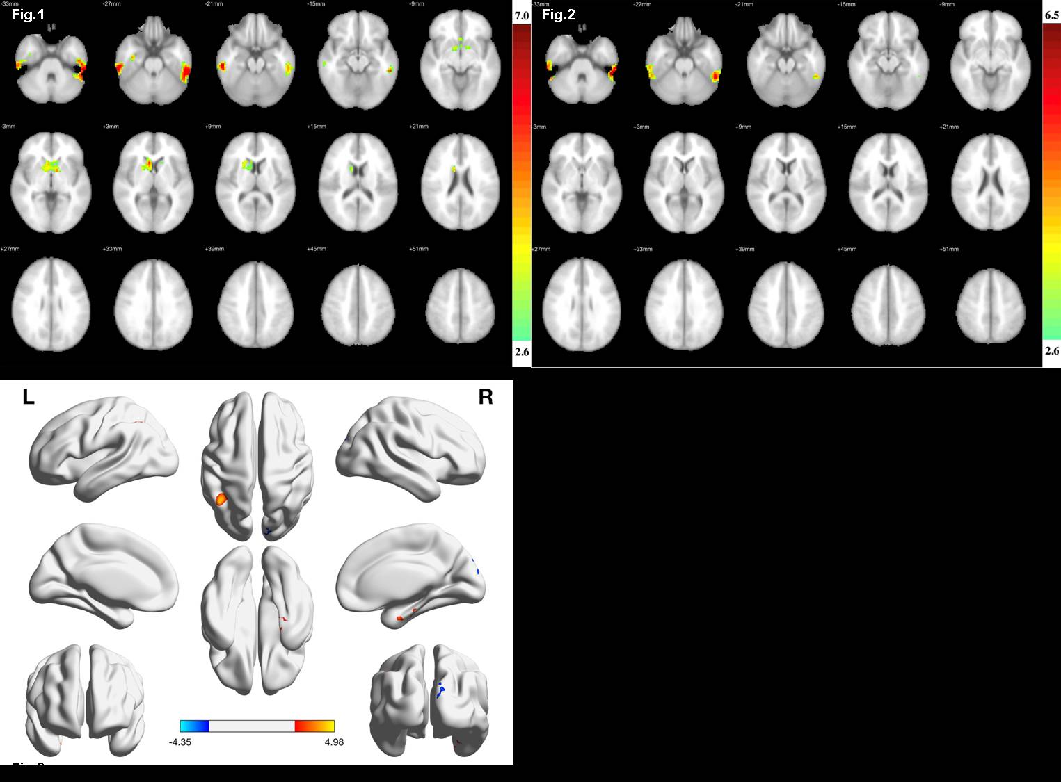
Fig. 1 Cortical regions revealed
significant differences of ReHo values between two groups (GRF corrected
p<0.05). The colorbar represents Z values. Fig. 2 Cortical regions
revealed significant differences of ALFF values between two groups (GRF corrected
p<0.05). The colorbar represents Z values.Fig. 3 The significant between group
differences of functional connectivities using bilateral inferior temporal
gyrus as seed.