1786
Clinical Equivalence Assessment of T2 Synthetic Pediatric Brain MRI1Pediatric Radiology, CHRU de Tours, Tours, France, 2Advanced Clinical Imaging Technology, Siemens Healthcare AG, Lausanne, Switzerland, 3Pediatric Radiology, CHRU de Tours, Tours, Switzerland, 4Pediatric Radiology, CHRU Tours, Tours, France
Synopsis
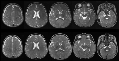
In a prospective randomized study, we compared the image quality of a synthetized T2 with conventional turbo spin echo T2 during pediatric brain MRI. According to several assessment criteria, synthetic T2 seemed to be an overall equivalent to standard TSE T2, with the advantage of new available T2 quantitative data with a similar acquisition time.
Introduction
Synthetic MR imaging provides different image contrasts as well as quantitative information from one scan [1]. It would well suit pediatric neuroradiology for various applications such as analysis of white matter disorders. However, synthetic contrasts have been poorly evaluated in this population [2]. The purpose of this study was to compare the image quality of synthetic T2 with conventional T2 turbo spin echo (TSE) in pediatric brain MRI.Methods
This was a mono-center prospective study. Synthetic and conventional MRI acquisitions at 1.5 Tesla (MAGNETOM Aera, Siemens Healthcare, Erlangen, Germany) were performed for each patient during the same session using a prototype accelerated T2-mapping sequence package (TAsynthetic=3:07 min, TAconventional=2:33 min). Image sets were blindly and randomly analyzed by senior and fellow pediatric neuroradiologists. Global image quality, morphologic legibility of standard structures and artifacts were assessed using a 4-point Likert scale. Inter-observer kappa agreements were calculated. The capability of the synthesized contrasts and conventional T2 TSE to discern normal and pathologic cases was evaluated.Results
Sixty patients were enrolled. The overall diagnostic quality of the synthesized contrasts was non-inferior to conventional imaging (p=0.06). There was no significant difference in the legibility of normal and pathological anatomic structures of synthetic and conventional TSE T2 (all p > 0.05) as well as for artifacts except for phase encoding (p=0.008). Interobserver agreement was good to almost perfect (kappa between 0.66 and 1).Discussion
Synthetic T2 contrasts seem to be an overall equivalent to standard TSE T2 for clinical practice in pediatric neuroradiology, after awareness of the rise of some artifacts. Those artifacts were easily recognizable, very similar from one patient to another, and differ considerable from anatomy not to be confounded with pathological conditions. The major benefit of this technique is to provide quantitative information on the inherent tissue relaxation. That could be used to build normative databases of healthy subjects which may be helpful to assess white matter myelination and maturation.
Conclusion
Quantitative T2 mapping together with synthesized T2 contrasts with the potential to build normative databases of healthy subjects, were found to provide clinically equivalent information in pediatric neuro-imaging compared to conventional T2 TSE sequences.Acknowledgements
The authors thank John Sheath for English langage assistance.References
1 Tanenbaum LN, Tsiouris AJ, Johnson AN, et al. Synthetic MRI for Clinical Neuroimaging: Results of the Magnetic Resonance Image Compilation (MAGiC) Prospective, Multicenter, Multireader Trial. AJNR Am J Neuroradiol. April 2017. doi:10.3174/ajnr.A5227. 7.
2 Betts AM, Leach JL, Jones BV, Zhang B, Serai S. Brain imaging with synthetic MR in children: clinical quality assessment. Neuroradiology. 2016;58(10):1017-1026. doi:10.1007/s00234-016-1723-9
