1770

T2 relaxometry MRI predicts cerebral palsy in preterm infants
Yi-Shan Tsai1, Li-Wen Chen2, and Feng-Mao Chiu3

1Department of Diagnostic Radiology, National Cheng Kung University Hospital, College of Medicine, National Cheng Kung University, Tainan, Taiwan, 2Departments of Pediatrics, National Cheng Kung University Hospital, College of Medicine, National Cheng Kung University, Tainan, Taiwan, 3Clinical MR application, Philips Healthcare, Taipei, Taiwan

Synopsis

T2 relaxometry brain MRI could be of prognostic value in preterm infants. The maturation patterns of periventricular white matter differed according to neurodevelopmental outcomes. T2 relaxation values over mid-body periventricular white matter at > 1 month old of corrected age could predict CP. T2 relaxometry brain MRI provides neuroimaging-outcome correlation among preterm infants, especially when interpreted with age-specific and area-selective considerations.

E-Poster (Electronic poster)

Introduction: T2 relaxometry brain MRI enables objective measurement of brain maturation based on the water-macromolecule ratio in white matter, but the outcome correlation is not established in preterm infants. Our study aims to predict neurodevelopment with T2 relaxation values of brain MRI among preterm infants. Methods: During January 1st 2012 to May 31st 2015, preterm infants who underwent both T2 relaxometry brain MRI and neurodevelopmental follow-up were retrospectively reviewed. T2 relaxation values were measured over periventricular white matter, including sections through frontal horns, mid-body of lateral ventricles, and centrum semiovale (Figure 1). Periventricular T2 relaxometry in relation to corrected age was analyzed with restricted cubic spline regression. Prediction of cerebral palsy was examined with receiver operating characteristic curve (ROC). Results: Thirty-eight preterm infants were enrolled for analysis (Figure 2). Twenty patients (52.6%) had neurodevelopmental abnormalities, including 8 (21%) of developmental delay without cerebral palsy, and 12 (31.6%) of cerebral palsy. The periventricular T2 relaxation values in relation to age was curvilinear in normal-developmental preterm infants, linear in developmental delay without cerebral palsy, and flat in cerebral palsy (Figure 3). When MRI was performed at > 1 month old of corrected age, cerebral palsy could be predicted with T2 relaxometry of periventricular white matter on sections through mid-body of lateral ventricles (Figure 4, area under the ROC = 0.738; cut-off value of >217.4 with 63.6% of sensitivity and 100.0% of specificity). Discussion: With the advances of neonatal intensive care, the macroscopic cystic periventricular leukomalacia are substituted by other forms of white matter lesions, including microscopic necrosis and diffuse white matter injury, both of which are prone to be underestimated in conventional qualitative MRI.1 T2 relaxometry is a fast scan to quantify tissue characteristics, providing an objective measurement of watery contents in brain.2 Our study found that white matter maturation patterns were different depending on the neurological outcomes, as T2 relaxometry showed curvilinear decline in normal-developmental preterm infants, linear decline in developmental delay, and flat in cerebral palsy. In our study of CP infants, the damaged WM revealed lower T2 relaxation than the normal watery WM in neonatal period, and higher T2 relaxation values than the normal myelinated WM in later infancy (Figure 5). In our observation, T2 relaxation values of mid-body periventricular white matter could predict CP when the MRI was performed after 1 month old of corrected age. In preterm infants, T2 relaxometry of >217.4 predicted CP with 63.6% of sensitivity and 100.0% of specificity. Conclusion: T2 relaxometry brain MRI could provide prognostic prediction on neurodevelopmental outcomes in premature infants. Age-dependent and area-selective interpretation in preterm brains should be emphasized.

Acknowledgements

We would like to thank Mr. Feng-Mao Chiu, the clinical MR application specialist of Philips Healthcare, for the support of simulation model in T2 estimation accuracy.

References

1. Back SA. Brain Injury in the Preterm Infant: New Horizons for Pathogenesis and Prevention. Pediatr Neurol 2015;53:185-92 2. He L, Parikh NA. Atlas-guided quantification of white matter signal abnormalities on term-equivalent age MRI in very preterm infants: findings predict language and cognitive development at two years of age. PLoS One 2013;8:e85475

Figures

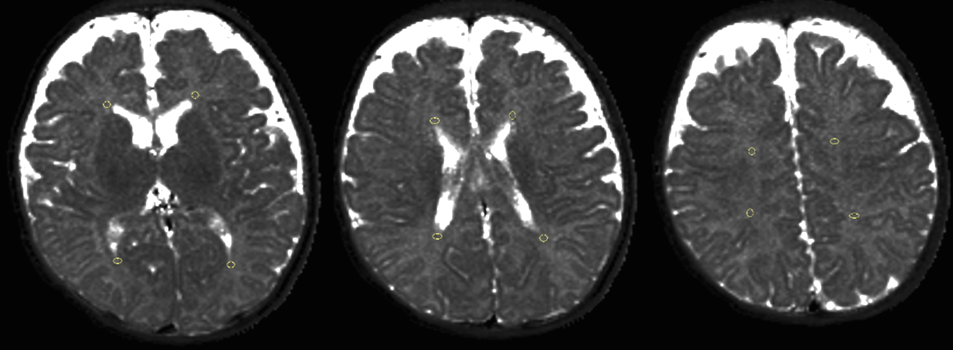
Figure 1. Manually drawn periventricular white matter on three axial sections through frontal horns, mid-body of lateral ventricles, and centrum semiovale. A circular ROI area is 5-8mm2. The four T2-mapping values of each section are averaged for calculation.

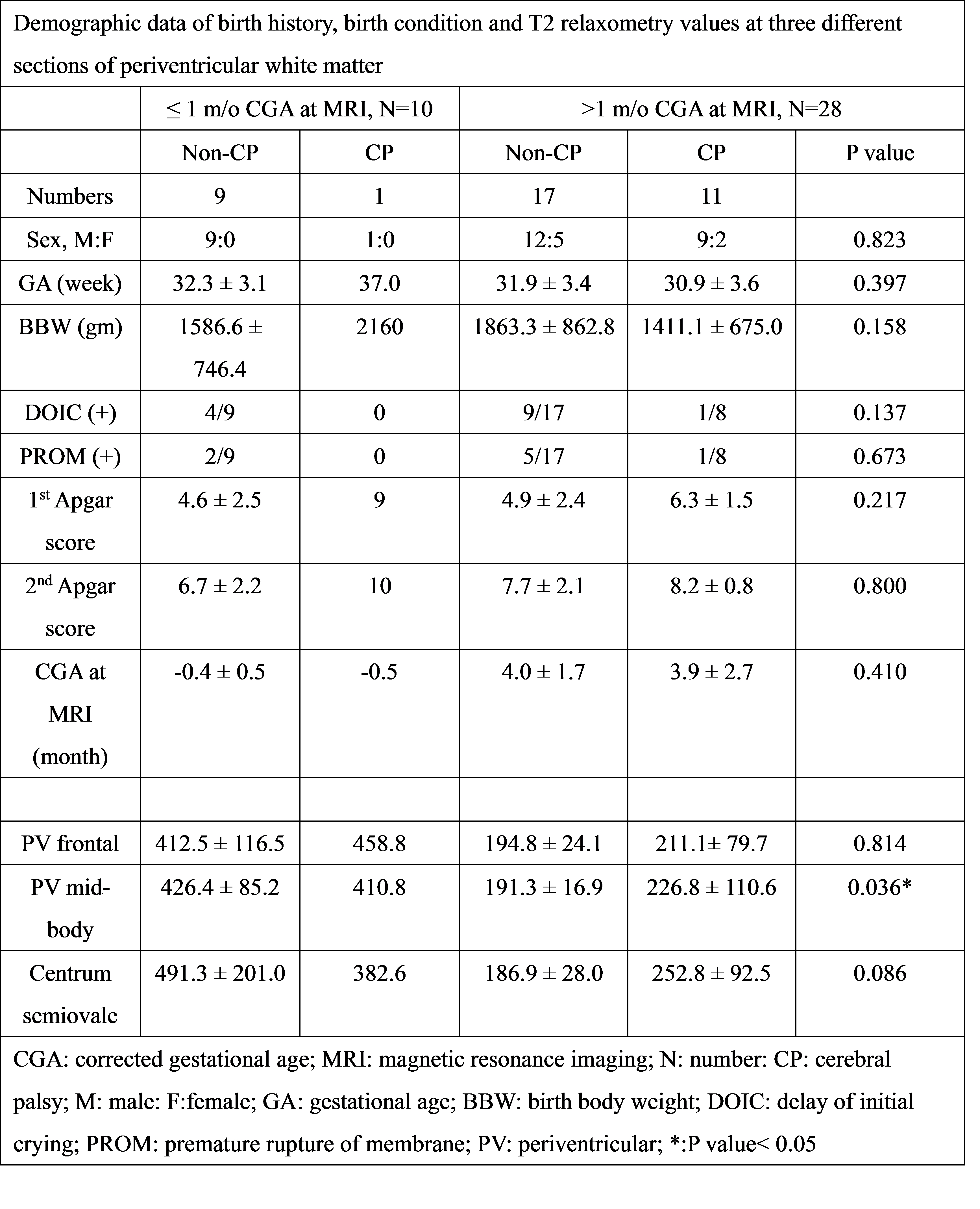
Figure 2. Demographic data of birth history, birth condition and T2 relaxometry values at three different sections of periventricular white matter

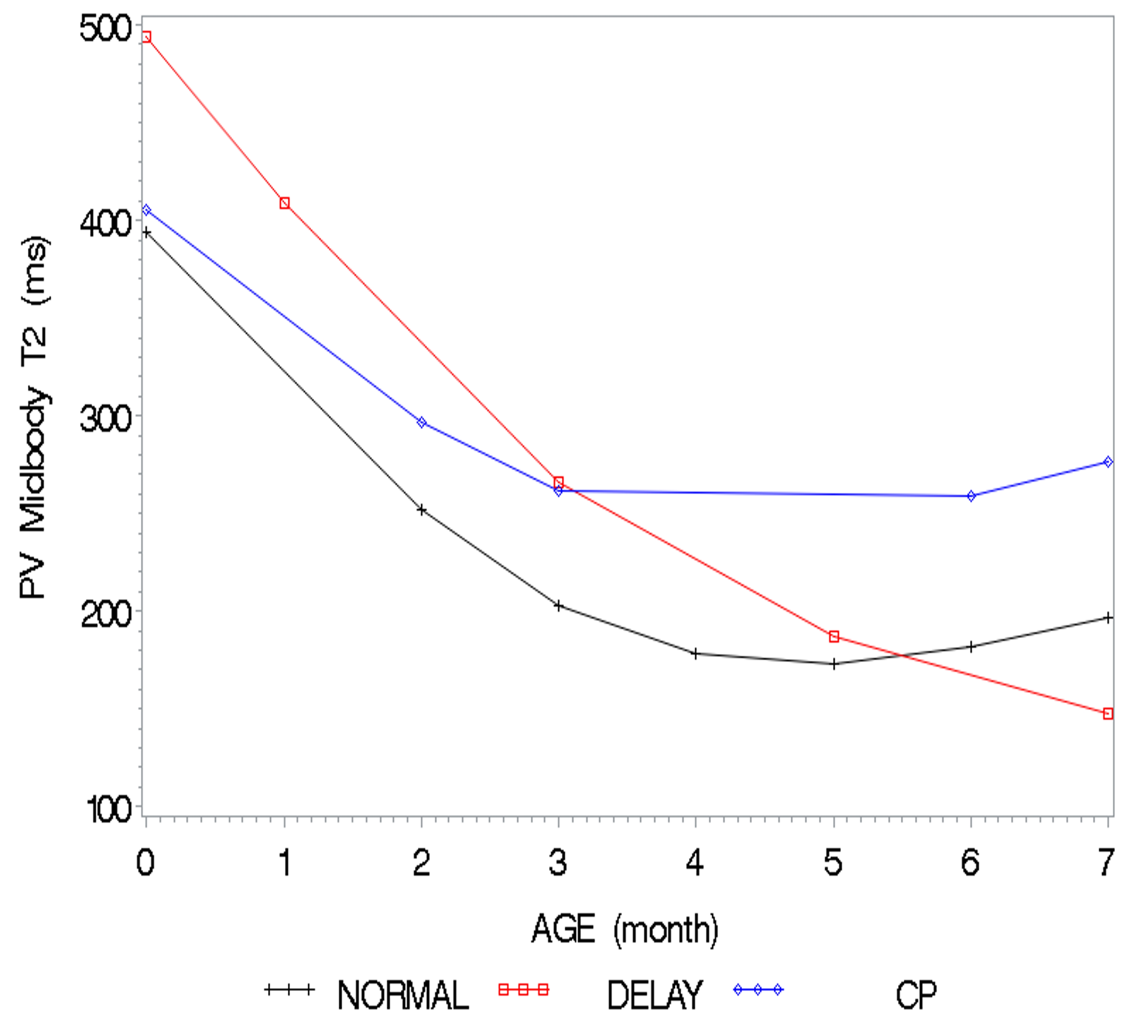
Figure 3. Restricted cubic spline regression analysis of T2 relaxometry versus age over mid-body periventricular white matter. T2 quantification values in relation to age are curvilinear, linear, and flat among normal development (black line), delayed development (red line), and cerebral palsy (blue line) preterm infants respectively.

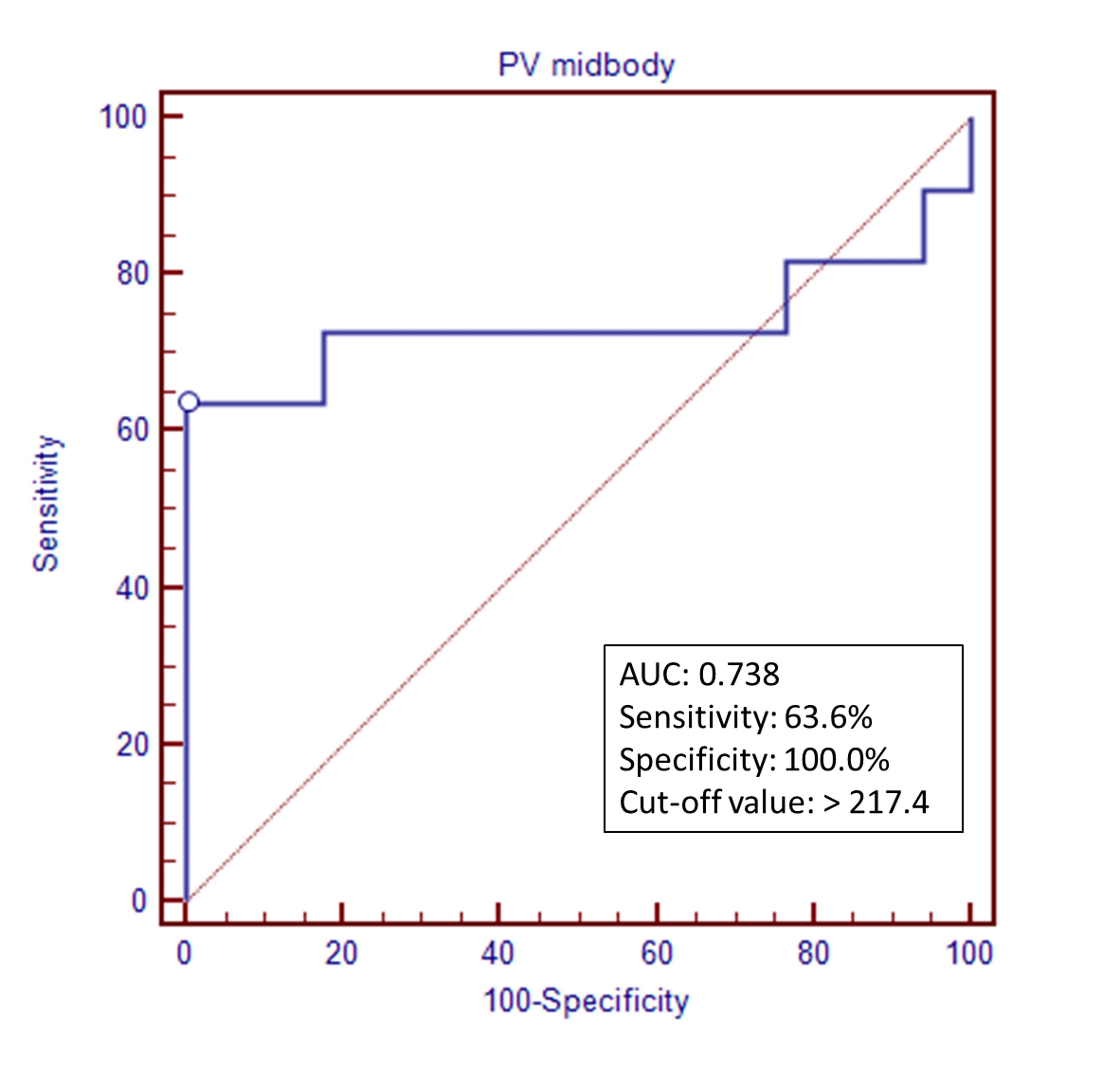
Figure 4. Receiver operating characteristic (ROC) curves of T2 relaxometry on the sections through mid-body of lateral ventricles.

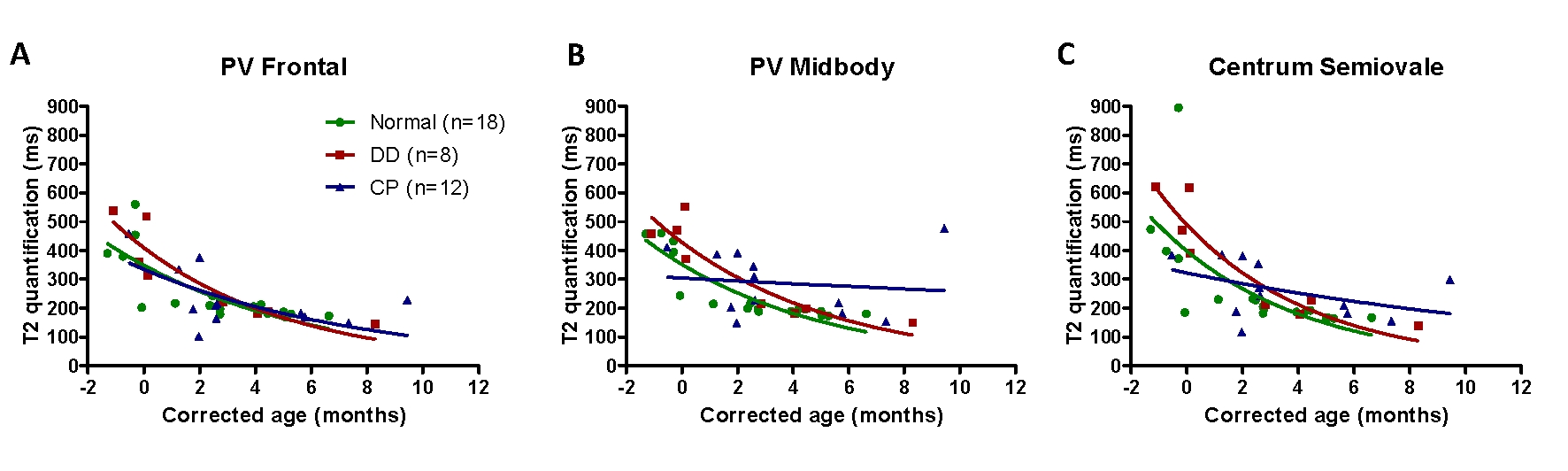
Figure 5. T2 relaxation values versus age among normal development, delayed development without cerebral palsy, and cerebral palsy patients. T2 quantification over periventricular white matter declines with age. Periventricular white matter through sections of (A) Frontal horns of lateral ventricles (B) Mid-body of lateral ventricles, and (C) Centrum semiovale. (CP: cerebral palsy; DD: delayed development (without cerebral palsy); PV: periventricular)

Proc. Intl. Soc. Mag. Reson. Med. 26 (2018)
1770