1765
Towards a high-resolution MRI Atlas of the Human Foetus: a Post-Mortem Pilot Study of ex-vivo preserved Foetal specimens at 7 Tesla.1Neurosurgery, University of Minnesota, Minneapolis, MN, United States, 2Integrative Biology and Physiology, University of Minnesota, Minneapolis, MN, United States, 3Radiology, University of Minnesota, Minneapolis, MN, United States
Synopsis
In an effort to expand the existing MRI reference material available to Medical professionals, including developmental anatomists,
Introduction
Methods
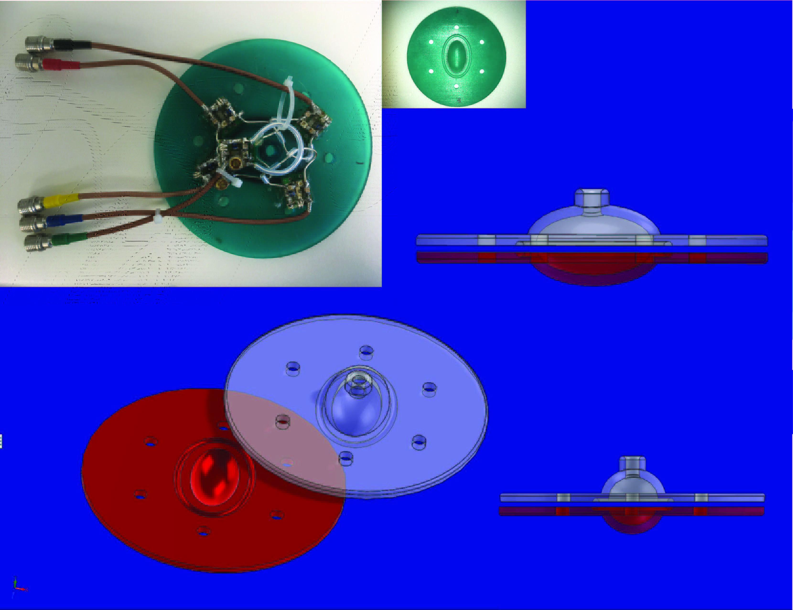
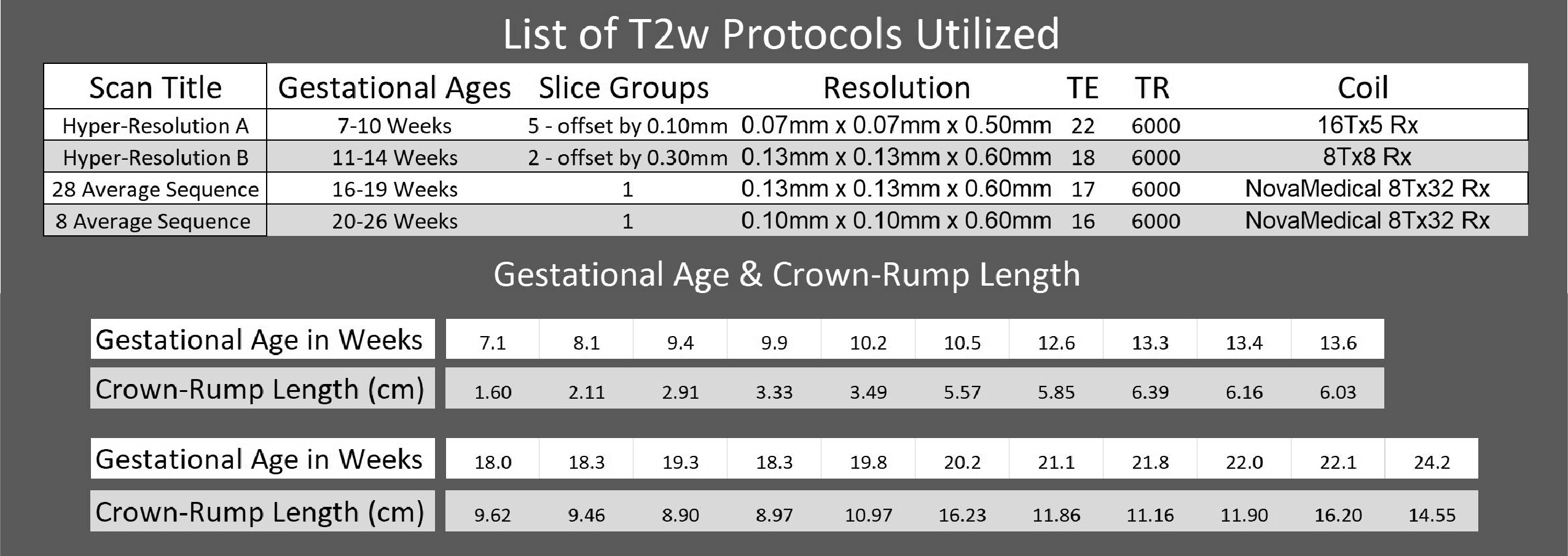
Workflow: CT scans of 21 specimens were first acquired at a resolution of 0.60mm. Thereafter, 3D volume reconstruction of CT data was performed with the use of Mimics Research, 3-matic Research and Autodesk Maya to design optimized individual specimen canisters for different age groups. These canisters were then printed with stereolithography printers using “Watershed XC1112” and “Formlabs Tough Resin”. Thereafter, the canisters were used to hold the specimens for scanning and the space around the specimens was filled with a medium devoid of 1H MR signal (Fomblin Y L-VAC 25/6) to achieve susceptibility matching and avoid artifacts. 3 separate coils were utilized for scanning, depending on the specimen size: (1) 7T NovaMedical 8Tx32 Rx, (2) Custom built 7T 8Tx8 Rx, and (3) Custom built 7T 16Tx5 Rx coils. A total of 4 specialized T2w sequences were developed and used for scanning as described below. The images were then segmented and reconstructed for viewing and analysis.MR scans
Initial pilot scans (not shown) showed stronger T2 than T1 or proton density contrast between tissues in these specimens. Therefore, subsequently, only T2 weighted imaging was performed. A clear superiority in conspicuity and sharpness dictated conventional spin-echo rather than turbo spin-echo, likely due to the short T2 relaxation of these preserved specimens. Due to technical limitations, the slice thickness was ≥0.5mm. Subsequently, in the smallest samples, where in-plane spatial resolution is the highest, smooth interpolation along the slice direction was obtained by collecting multiple data sets shifted by a fraction of slice thickness.Results
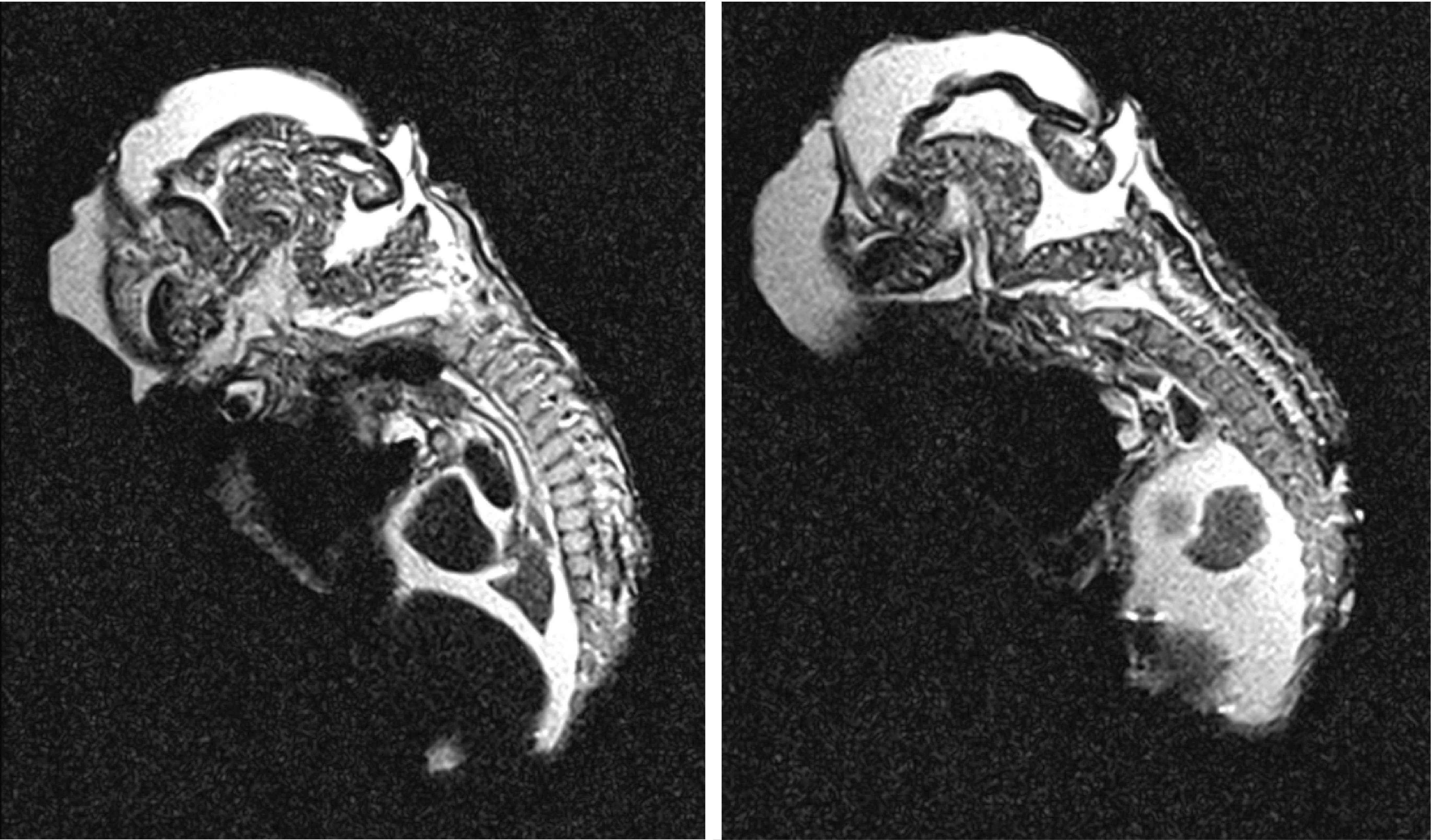
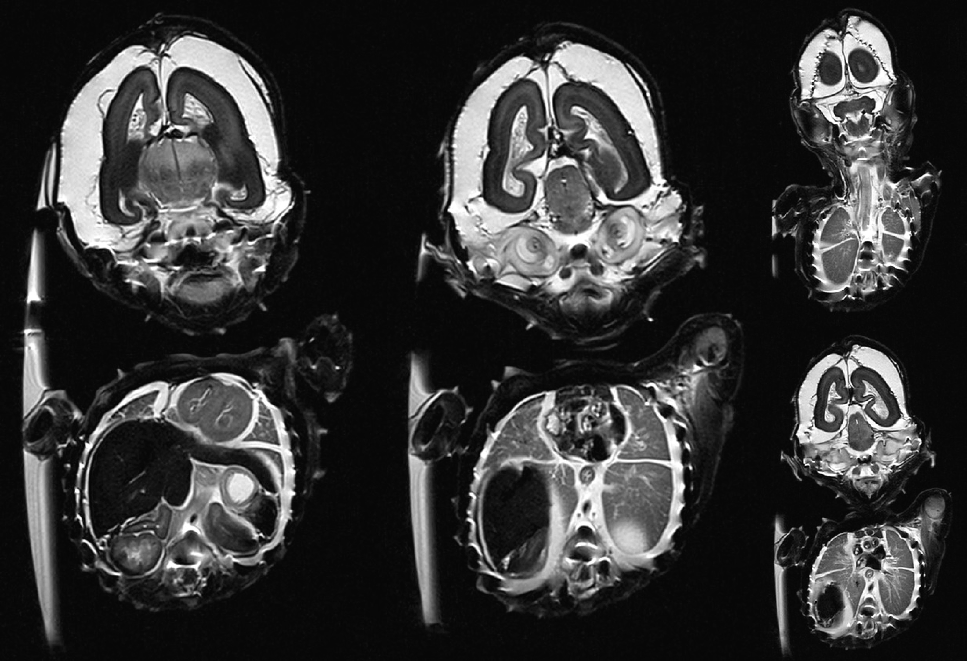
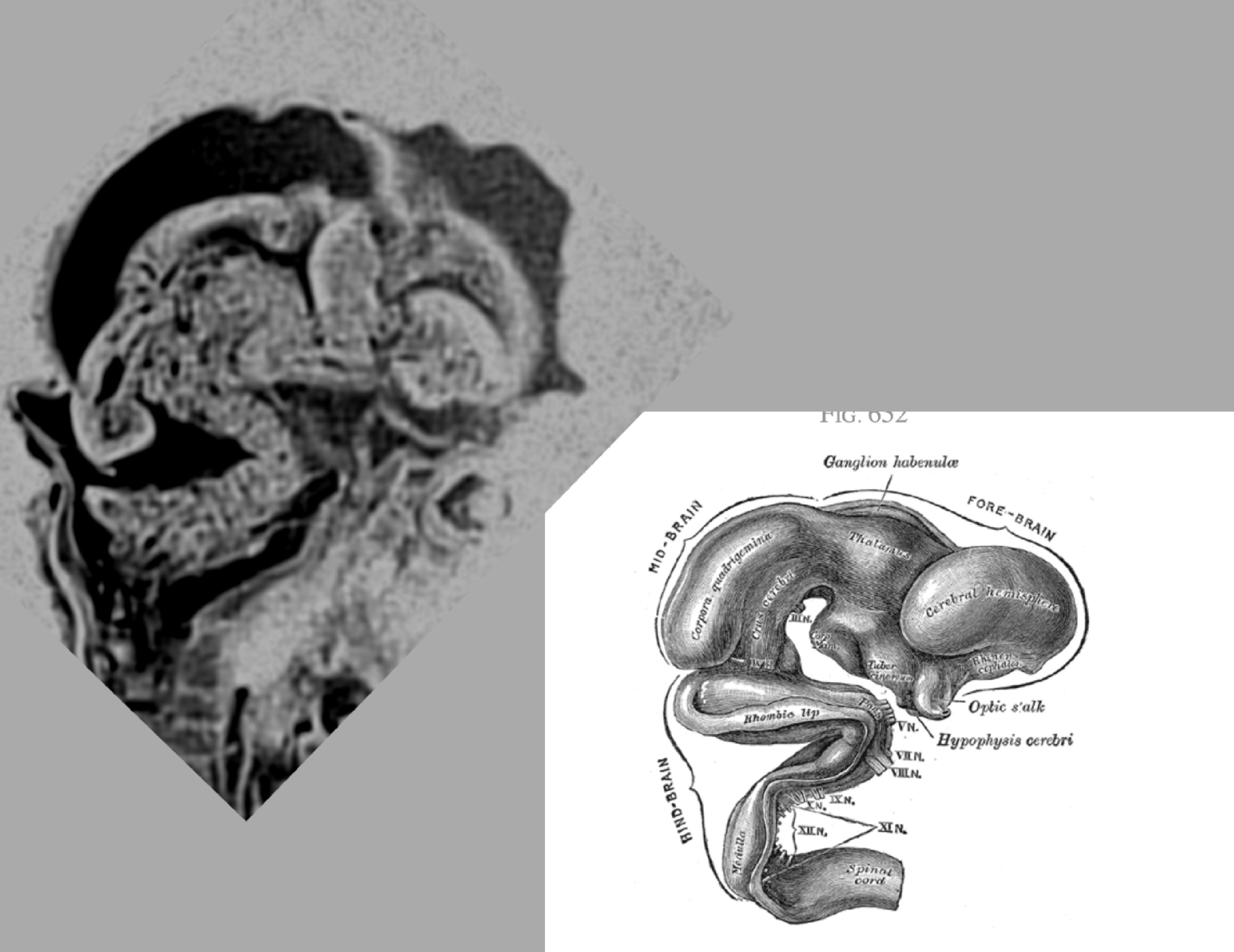
Optimizing SNR required building 4 different canisters to adapt with sample size. A striking characteristic observed throughout the study was the persistence of strong T2 contrast in all specimens. The Field of view and spatial resolution needed specific tradeoffs for the range of 21 different sample sizes while satisfying inherent matrix size max limits of our system, yielding 4 T2w protocols for optimal imaging, summarized in Table 1. High contrast and high resolution images of different organ systems, especially the Central Nervous system, the cardiovascular system and visceral organs could be reliably obtained even in the smallest specimens. For instance, the migration of cortical neurons from the peri-ventricular region to the cortical surface could be reliably demonstrated in the specimens of different gestational ages. Likewise, the folding of the neuronal tube and the folding of the heart chambers could be demonstrated reliably.Discussion & Conclusion
Due to the presence of a stable T2 contrast in all examples, general body imaging produced good results in these small specimens of short gestational age. Imaging of similar specimens at ultra high field strengths has not been previously documented1,2. This new collection of data will serve to produce additional reference texts that will extend the available resources for medical professionals, as well as further our understanding of organ development.Acknowledgements
P30 NS076408
P41 EB015894
References
1. Griffiths, Paul D., et al. Atlas of Fetal and Infant Brain MR. Elsevier Health Sciences, 2009.
2. Garel, C. Mri of the Fetal Brain: Normal Development and Cerebral Pathologies. Springer, 2013.
3. Gray, Henry. Anatomy of the Human Body. Philadelphia: Lea & Febiger, 1918; Bartleby.com, 2000
Figures