1758
A Bo Tapestry: MRI Magnet Technology, 1977-20171Upstate Medical University, Syracuse, NY, United States, 2Medical University of Vienna, Vienna, Austria, 3University of Sheffield, Sheffield, United Kingdom, 4Lakeside Imaging-e, Erlangen, Germany
Synopsis
This is a preliminary report from a project to gather and organize an objective historical record of human MRI scanner technology. This report spans magnet technology from 1977 to present (2017), covering about 100 magnets and scanners, and invites additional information.
Introduction & Purpose
This is a preliminary report from a project initiated with intent to gather, organize, and share an objective, verified historical record of MRI technology. This history includes contributions from inventors and “makers” in industry that are seminal to the evolution of MRI, but are largely silent or unfamiliar to many in the current ISMRM community. The aims of this preliminary report are (a) to present the data collected to date, especially the technology associated with Bo magnets; and (b) to solicit information and advice from other sources, especially documents and persons not yet familiar to these authors.Methods
We created a spreadsheet database of past and current scanners, tabulating technology characteristics and other attributes chosen as being critically important to current prevailing applications of MRI. The scope and content of this database was inspired and populated from old and recent publications, and the personal libraries and memories of the authors and a few others interviewed for this purpose.Results
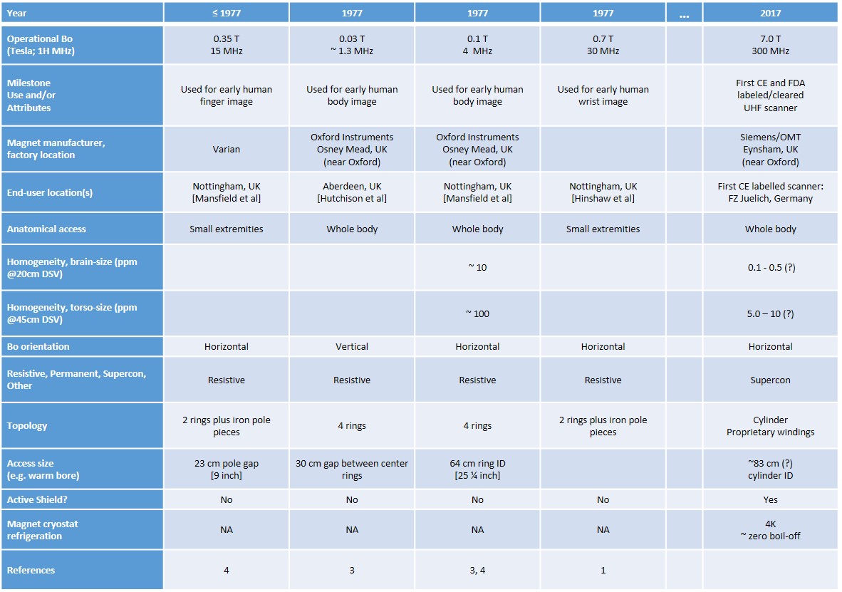
The database currently includes about 100 scanners, and 100 magnets, over the time interval from 1977 to 2017. Some characteristics of magnets (and other scanner components) are definitively described in published reviewed literature, but many are not. Some representative content of the database, showing a few magnets for scanners built at the chronological extremes of 1977 and 2017, is shown in the table in Figure 1.Discussion and Conclusions
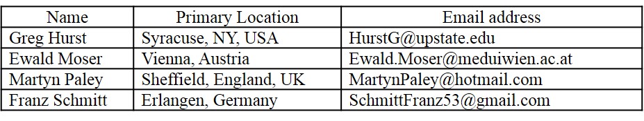
We believe that much additional relevant information, especially for early scanners, is held in personal archives and memories, and therefore will be practically available for a limited time. Accordingly, additional information for this database, and related comments, are highly welcome, and can be sent to any of the authors, using contact information given in the table in Figure 2.Acknowledgements
No acknowledgement found.References
1. Hinshaw WS, Bottomley PA, Holland GN. Radiographic thin-section image of the human wrist by nuclear magnetic resonance. Nature 1977.
2. Witkofksi RL, Karstaedt MB, Partain CL (editors). NMR Imaging: Proceedings of an International Symposium on Nuclear Magnetic Resonance Imaging, Bowman Gray School of Medicine. 1981.
3. Wood A. Magnetic Venture: The Story of Oxford Instruments. Oxford U Press. 2001.
4. Morris P. Whole-body MRI in Nottingham: The First Steps. Encyclopedia of MR. 2012.
5. Moser E, Laistler E, Schmitt F, Kontaxis F. Ultra-High Field NMR and MRI – The Role of Magnet Technology to Increase Sensitivity and Specificity. Front. Phys. 2017 August; Vol 5, article 33.
Figures