1755
A feasibility study of ultra-high-strength gradient system on 3T: demonstration using DTI on anisotropic diffusion fibre phantoms1Institute of Biomedical Engineering and Nanomedicine, National Health Research Institutes, Miaoli, Taiwan, 2Institute of Neuroscience and Medicine – 4, Forschungszentrum Juelich, Juelich, Germany, Juelich, Germany, 3Department of Neurology, Faculty of Medicine, RWTH Aachen University, Aachen, Germany
Synopsis
In this study, we aimed to integrate an ultra-high-strength gradient system (15 gauss/cm) on a 3T scanner and to demonstrate its feasibility by employing diffusion tensor imaging (DTI) on dedicated anisotropic diffusion fibre phantoms. Two DTI experiments were performed to explore the feasibility of this gradient system, i.e. comparisons of gradient strengths and number of averages. Our results demonstrate reasonable SNR and diffusion contrast acquired on this system using pulsed gradient spin echo diffusion weighted scans could provide useful information. Consistently, it also suggests higher gradient strength could be beneficial to improve the quality of diffusion MRI experiments and its ability to resolve fibre orientations, especially when higher b-values are used.
Purpose
The recent advance in MRI hardware, especially the ultra-high-strength gradients, has shown their potential to decipher the complex brain structures and their associated connectivity. The higher gradient strength up to 30 gauss/cm has been demonstrated to provide superior angular resolution to map brain connections in vivo by using diffusion MRI techniques [1,2]. Accessibility of this on clinical scanners is, however, specifically limited by, for example, hardware demand and high cost. In this study, we aimed to integrate an ultra-high-strength gradient system (15 gauss/cm) on a whole-body 3T magnet and to demonstrate its feasibility by employing diffusion tensor imaging (DTI) on dedicated anisotropic diffusion fibre phantoms.Methods
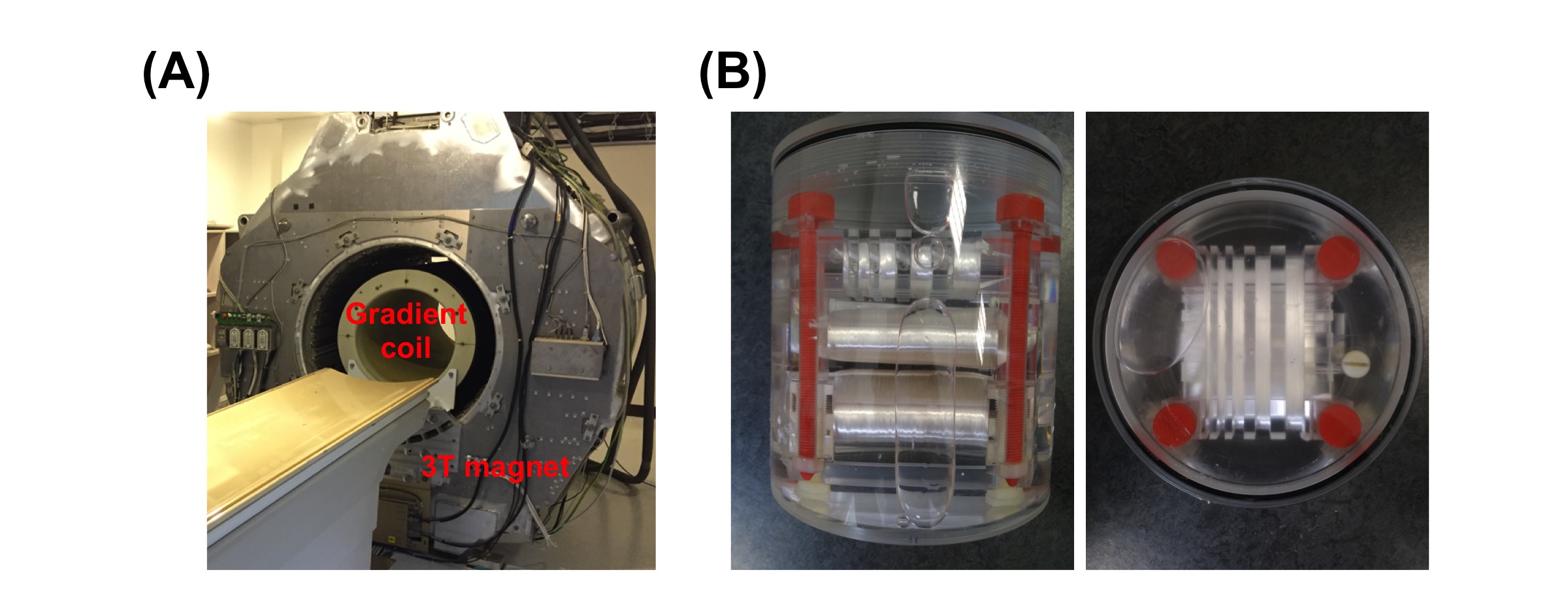
In this work, an ultra-high-strength gradient coil has been integrated on a 3T whole-body magnet (15 gauss/cm, Resonance Research Inc., Billerica, MA, USA) [3]. A gradient power amplifier (Analogic, Peabody, MA, USA) was calibrated and matched to drive the gradient coil. The MRI system and all the sequences were controlled by a spectrometer operated at 127.7 MHz (MR Solutions Ltd., Surrey, UK). The RF transmission and signal reception were achieved by using a custom-made birdcage coil with an inner diameter of 26 cm. The system overview and dedicated fibre phantoms are shown in figure 1. Two sets of DTI experiments were performed in order to explore the feasibility of this gradient system, i.e. comparisons of gradient strengths (exp#1, 4 vs. 8 gauss/cm) and number of averages (exp#2, NEX of 1~9). The dedicated home-built phantoms [4] were measured using a pulsed gradient spin-echo (PGSE) diffusion-weighted (DW) imaging sequence. In exp#1, sequence parameters for the lower gradient strength (4 gauss/cm) were TR of 2000 ms, Δ of 35 ms, field-of-view (FOV) of 200x200 mm2, matrix size of 128x128, slice thickness of 2 mm, 5 slices and maximum b-values (bmax) of 330/800/1120 s/mm2, corresponding to TE of 61/67/71 ms and δ of 10/16/20 ms, respectively. With the same spatial resolution, the sequence parameters for higher gradient strength (8 gauss/cm) were TR of 2000 ms, Δ of 23 ms and bmax of 330/860/1190 s/mm2, corresponding to TE of 45/49/52 ms and δ of 6/10/12 ms, respectively. For exp#2, the sequence parameters are TR/TE of 4000/49 ms, Δ/δ of 23/10 ms, FOV of 192x192 mm2, matrix size of 96x96, slice thickness of 2 mm, 17 slices and bmax of 860 s/mm2. All DTI reconstruction processes were performed using an in-house program written in C language, and the reconstructed DTI indices (MD: mean diffusivity, FA: fractional anisotropy and cFA: colored-coded FA) were visualized using ImageJ software.Results
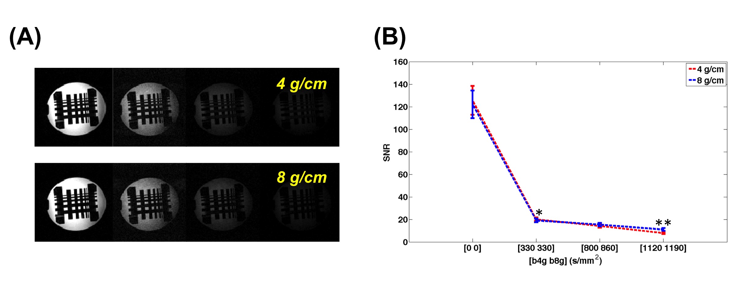
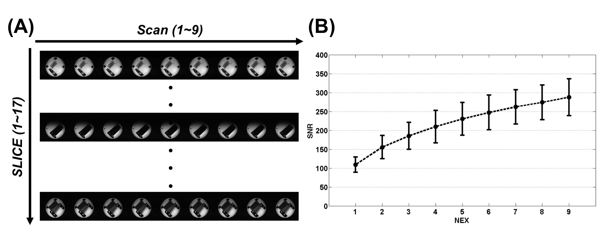
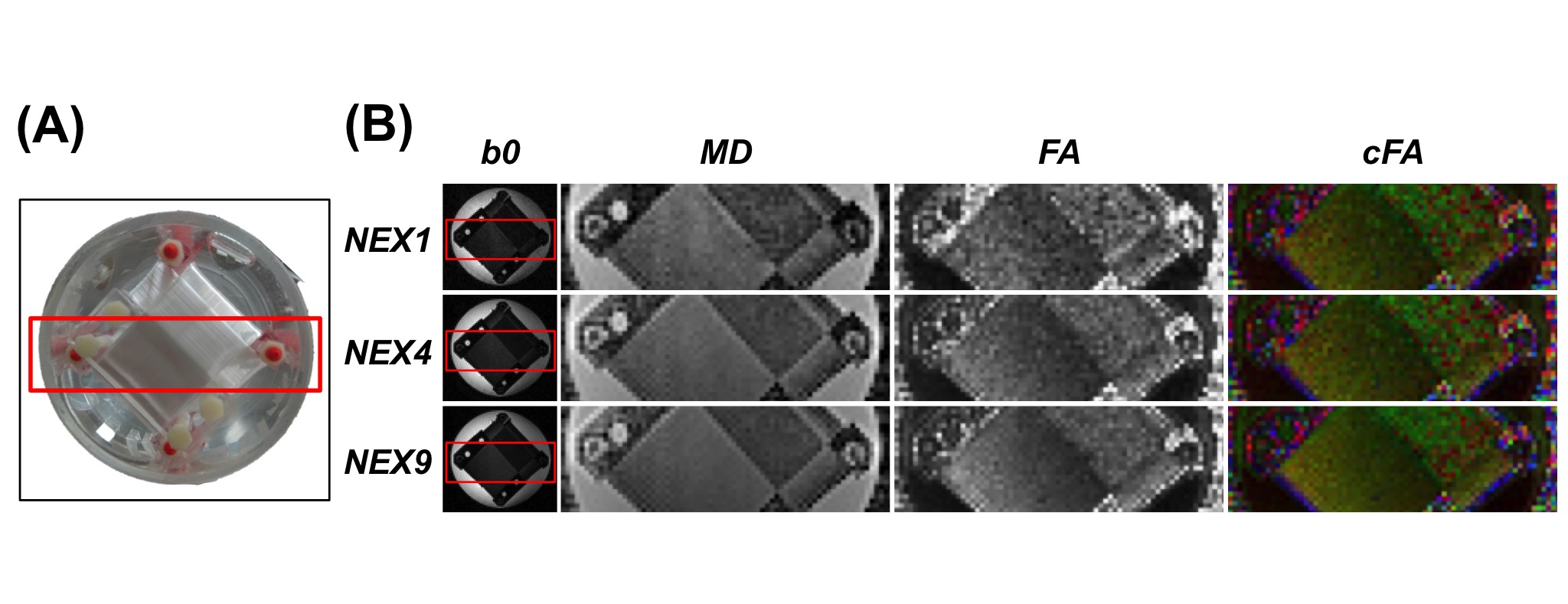
Figure 2A shows the comparison of DW images acquired by two different gradient strengths, i.e. 4 and 8 gauss/cm. The calculated signal-to-noise ratios (SNRs) on each b0 and DWI were averaged among all slices for statistical comparison between two gradient strengths. The SNR values are plotted against bmax for both gradient strengths are shown in figure 2B confirming that employing higher gradient strength could significantly improve SNR values (~1.5 folds by comparing the highest bmax in both gradient settings). Figure 3A shows all b0 images (each separate scan before averaging) acquired in exp#2 by employing the higher gradient strength (8 gauss/cm). The SNR for different NEX (1~9) was calculated by averaging different number of scans. The SNR values averaged from all slices are plotted in figure 3B, showing an increasing tendency as NEX increases. This result demonstrates that our system could provide minimally acceptable SNR values and diffusion contrast for PGSE DW scans. Figure 4A and 4B show the imaging plane of fibre phantom and its corresponding b0, MD, FA and cFA images reconstructed from DTI datasets with NEX of 1, 4 and 9, respectively. Consistent with results of SNR comparison, the DTI indices, i.e. MD and FA, reconstructed from data with NEX of 9 are less noisy and could provide better accuracy in resolving fibre orientations in homogeneous region as revealed in cFA mapping.Discussion and Conclusions
In this study, we have successfully integrated an ultra-high-strength gradient system on a whole-body 3T magnet. Its feasibility on DTI has also been demonstrated by assessing the SNR of b0 images and its relationship with gradient strengths and NEX. Our results also suggest that higher gradient strength could be beneficial to improve the quality of diffusion MRI and its ability to resolve fibre orientations, especially when higher b-values are used.Acknowledgements
This study is supported by NHRI intramural grants (NHRI-BN-PP-06, NHRI-BN-SP-01 and NHRI-BN-SP-05) and MOST-DAAD grant 106-2911-I-400-502. We also thank for Dr. Piotr Starewicz of RRI for his valuable consultation and technical support.References
[1] Setsompop et al., Neuroimage, 2013, 80, p220-33
[2] McNab et al., Neuroimage, 2013, 80, p234-45
[3] Chen et al., ESMRMb 2017, p264
[4] Farrher et al., MRI, 2012, 30, p518-526
Figures