1752
Calibration of Siemens MAGNETOM(TM) Terra 7T Shim System and Analysis of Static 3rd-order B0-Shimming of the Heart Using B0DETOX1Chair of Cellular and Molecular Imaging, Comprehensive Heart Failure Center (CHFC), University Hospital, Wuerzburg, Germany, 2Translational Center Regenerative Therapies (TLC-RT), Fraunhofer Institute for Silicate Research (ISC), Wuerzburg, Germany, 3Departments of Biomedical Engineering and Radiology, Columbia University, New York, NY, United States
Synopsis
Susceptibility-induced field inhomogeneities in both space and time make B0-shimming a prerequisite for cardiac MRI at ultra-high field. All individual terms of the static 3rd-order spherical harmonics shim system were calibrated. Field mapping and calculation of shim currents are performed in customized B0DETOX software. Analysis of B0-inhomogeneities is later tested both in measurement of an ex-vivo pig heart and in-vivo in humans. The adjustment of the shim volume to the three measured slices in a healthy volunteer reduced the standard deviation of the field map by 4%, 19% and 18% compared to shimming of the global heart.
Introduction
Magnetic resonance imaging of the heart at ultra-high field (UHF) is challenging because of a variety of factors such as strong susceptibility-induced B0-field variations occur because of the complex structure of both the heart and surrounding tissue1,2. These variations result in a significant signal loss and in image distortions. Thus, their correction both spatially and temporally is an absolute prerequisite3. For the assessment of the spatial distribution of B0 in the volume of interest, the phase method is often used. Here, sufficient SNR at the longest TE is important for efficient phase-based B0-mapping. To analyze the magnetic field generated by spherical harmonics (SH) shim coils, the Siemens MAGNETOMTM Terra 7T 3rd-order SH shim system was first calibrated using phase-mapping pulse sequence for cardiac application. Customized B0DETOX4 software based on the calibration matrix later was used for field analysis in ex-vivo pig hearts as well as in several healthy volunteers.Methods
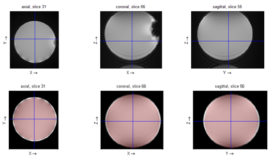
For 3rd-order shim calibration phase maps are acquired using a multi-slice gradient-echo pulse sequence (FOV 186×220×173 mm3, matrix size 108×128×64, TR=30 ms, FA=25°) measuring four single-TE scans (TE1/TE2/TE3/TE4 = 1.0/1.3/2.0/4.0 ms) for each shim setting with a 2-channel Tune-Up coil. B0-maps are calculated on pixel basis from the phase evolution over TE using linear regression. Spatial phase-unwrapping was performed using FSL5 algorithm. The magnetic field selected by region-of-interest (ROI) (Fig. 1) is decomposed by customized B0DETOX software into SH components taking into account that three 3rd-order terms (X3, XYZ and Y3) are not implemented in the 3rd-order shim electronics of the Terra system. To calibrate an individual shim term, its value was changed in seven steps relative to a reference B0-map using approximately 10 % of the dynamic range for the linear gradients, and 30 % for the higher orders. The calibration matrix A connects the shim settings s with all SH components f before shimming6, $$$s = -f·A^{-1}$$$ , and also yields information about the cross terms. B0DETOX-based field mapping is finally used to analyze localized scanner-side “Brain” shimming protocol which offers the shortest TE1 and ΔTE (TE1/TE2 = 1.02/3.06 ms)7. For both ex-vivo measurements of excised pig hearts and in-vivo measurements in healthy volunteers, a multi-slice gradient-echo pulse sequence is used (FOV 300×300×30 mm3, matrix 128×128×3, TR=100.0ms, TE1/TE2/TE3 = 1.70/2.83/3.96 ms). Ex-vivo measurements are performed with excised pig hearts preserved in 0.9 % NaCl solution. After granting permission by local ethics committee and obtaining informed consent from the volunteers, in-vivo measurements are performed under breath-hold using ECG-triggering. The improvement by a certain shim can e.g. be described by the quality factor QS = σR/σT which describes the decrease of the standard deviation σR of the resulting field compared to the initial field map σT.Results and Discussion
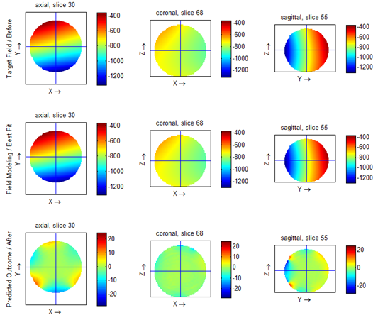
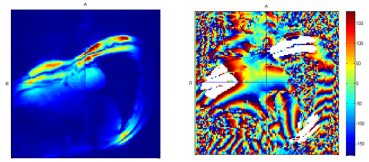
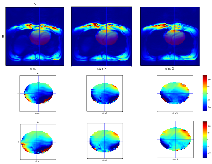
The SH shim system was successfully calibrated. The total dynamic ranges for each term are X = -2124 Hz/cm, Y = 2120 Hz/cm, Z = 2129 Hz/cm, X2-Y2 = 6.1 Hz/cm2, ZX = 12.6 Hz/cm2, Z2 = 40.2 Hz /cm2, ZY = -12.3 Hz/cm2, XY = -6.2 Hz/cm2, Z(X2-Y2) = -0.10 Hz/cm3, Z2X = -0.35 Hz/cm3, Z3 = -0.68 Hz/cm3, Z2Y = -0.34 Hz/cm3. The predicted residual field of the spherical phantom used for calibration shows the lacking 3rd-order in axial slice (Fig. 2). The quality of the prediction largely matches the experimental outcome. Analysis of short- and long-axis view images and field maps of the heart for a human participant are shown on Figures 3 and 4. Using 3rd-order shim calculation by B0DETOX in the heart, one has to take oblique slice orientation and resulting rotated coordinate system relative to the scanner’s gradient axes into account when acquiring short-axis or four-chamber view field maps. Scanner-side “Brain” shimming was both adjusted global on the heart and specifically on measured slices (Fig. 5). Quality factors QS for the three measured slices were QS,1=0.96, QS,2=0.81 and QS,3=0.82, which means an improvement in standard deviation of 4%, 19% and 18%.Conclusion
Calibration-based B0DETOX field analysis shows the potential for static 3rd-order SH B0-shimming optimization in both ex vivo and human measurements. However, further steps have to be done to perform proper in vivo shimming of the heart. These steps include dynamic shimming adjusted both on cardiac phases and slices as well as static and dynamic multi-coil shimming. In contrast to scanner-side shimming – which would require a very long acquisition time - B0DETOX enables calculation of field maps measured using ECG-triggering. Calculation can be performed for specific heart phases, which will further improve shim quality.Acknowledgements
Financial support was obtained from the German Ministry of Education and Research (BMBF) under grant #01E1O1504.References
[1] W. Mattar, C. Juchem, M. Terekhov, L.M. Schreiber; Multi-coil B0 shimming of the human heart: A theoretical assessment; ISMRM Annual Meeting, Singapore (2016)
[2] M.K. Atalay, B.P. Poncelet, H.L. Kantor, T.J. Brady, R.M. Weisskopf; Cardiac susceptibility artifacts arising from the heart lung interface; Magnetic Resonance in Medicine 45, 341-5 (2001)
[3] P.F. Ferreira, P.D. Gatehouse, R.H. Mohiaddin, D.N. Firmin; Cardiovascular magnetic resonance artefacts; Journal of Cardiovascular Magnetic Resonance, 15:41(2013)
[4] C. Juchem, S.U. Rudrapatna, T.W. Nixon, R.A. de Graaf; Dynamic multi-coil technique (DYNAMITE) shimming for echo-planar imaging of the human brain at 7 Tesla, NeuroImage 105 (2015), 462 - 472
[5] S.M. Smith, M. Jenkinson, M.W. Woolrich, C.F. Beckman, T.E.J. Behrens, H. Johansen-Berg, P.R. Bannister, M. De Luca, I. Drobnjak, D.E. Flitney, R. Niazy, J. Saunders, J. Vickers, Y. Zhang, N. De Stefano J.M. Brady, P.M. Matthews; Advances in functional and structural MR image analysis and implementation as FSL; NeuroImage, 23(S1): 208-19 (2004)
[6] C. Juchem, T.W. Nixon, P. Diduch, D.L. Rothman, P. Starewicz, R.A. de Graaf; Dynamic Shimming of the Human Brain at 7 T, Concepts in Magnetic Resonance Part B (Magnetic Resonance Engineering), Vol. 37B (3) 116 – 128 (2010)
[7] M. Hock, M. Terekhov, D. Lohr, A. Schröder, H. Walles, L.M. Schreiber; B0-Mapping and Shimming Efficiency for ex Vivo MR Imaging of the Heart at Ultra-High Field – Validation of Standard Shimming Protocols of MAGNETOMTM Terra 7 T Scanner; DGMP and DGBMT Annual Meeting, Dresden (2017)
Figures