1741
MR Compatibility of MADPET4: A Small Animal PET Insert for a 7T MRI System1Nuclear Medicine, Klinikum rechts der Isar, Technical University of Munich, Munich, Germany, 2Physics, Technical University of Munich, Garching, Germany, 3Nuclear Medicine, University Hospital of LMU Munich, Munich, Germany
Synopsis
The impacts of operating a small animal PET insert in a 7T MRI system were studied. The MRI's performance was compared with and without the insert by measuring the static field, flip angle distribution, RF noise, and several imaging sequences with two RF volume coils. With the insert inside a large 1H volume coil, the MR was limited to T1-weighted anatomical imaging, and required a surface receive coil for adequate SNR. With the insert enclosing a small 1H/13C volume coil, the primary impact on MRI was up to 38% reduced SNR, and all tested MRI sequences were functional.
INTRODUCTION
MADPET4 is a small animal PET insert designed to be operated inside a 7T Agilent/GE MR901 Magnet (currently operating with Bruker AVANCE III HD electronics)1. In this work, the impact of the PET insert on the MRI system is assessed by comparison of image quality, noise, and B0 and B1 fields, with several RF coil configurations at 1H and 13C frequencies, with and without the insert in the bore or acquiring PET data. These results are important to evaluate the suitability of the PET insert design for use in combined and simultaneous PET/MRI experiments.
METHODS
The MADPET4 insert has 2640 Ce:LYSO scintillation crystals, arranged in a dual layer configuration, individually read out by silicon photomultipliers (SiPMs), providing high count rate capability and depth of interaction correction. No RF shielding and no active electronic components are located in the MR scanner bore. Instead, 1.5 m coaxial cables connect the SiPMs to the readout ASIC boards located outside the bore, which are in turn connected by 3 m coaxial cables to the digital data acquisition electronics located outside the MR scanner room Faraday cage. The insert has an inner diameter of 88 mm and outer diameter of 150 mm.
The PET insert's impact on MRI was tested with a 1H-frequency “large” volume coil (surrounding the insert, inner diameter 150 mm), and with a dual-tuned 1H/13C-frequency “small” volume coil (within the insert, inner diameter 31 mm) (both from RAPID Biomedical). MR compatibility was assessed with 1H B0 static field mapping using the scanner's FieldMap dual gradient echo sequence, 1H B1 mapping by varying the excitation pulse power of a gradient echo sequence, 1H gradient echo imaging, 1H spin echo imaging, 1H spin echo and FID EPI imaging, 1H and 13C noise scans (wide bandwidth spectra acquired without using gradients or RF excitation), and 13C chemical shift imaging. All tests were compared between cases without the insert, with the insert in place but inactive, and with the insert acquiring.
RESULTS
With the PET insert in the large 1H volume coil, compared with no insert: B0 variations of +/- 1 ppm were observed on the periphery of the PET enclosed volume (Figure 1), corresponding to the positions of the cable connector on the PET insert's inner ring SiPM boards. B1 was reduced by approximately 62% (lower flip angle for a given excitation power) near the magnet and RF coil centre. In 1H noise spectra (Figure 2), noise increased with or without PET acquiring across all measured frequencies, and additionally at various narrow frequency spikes while PET was acquiring. Combined with the already-limited RF transmit performance of this coil with rat or mouse sized subjects (for which it is not normally used), operating with the PET insert required very low transmit bandwidth for 90 and 180 degree pulses (eg. 600 Hz).
With the 1H/13C small volume coil in the PET insert, compared with no insert: Negligible effects on B0 and B1 (Figure 3) were observed. In 1H and 13C noise spectra (Figure 2), 1H noise was unchanged when PET was not acquiring, but increased 30-50% across all measured frequencies and at numerous narrow frequency spikes while PET was acquiring, whereas 13C noise increased by more than double, regardless of whether PET was acquiring. In MR images (Figure 4) 1H MRI SNR was reduced by 23-30%, depending on the imaging sequence used, and 13C CSI SNR was reduced 38% while PET was acquiring.
DISCUSSION
With the large RF volume coil: B1 effects are largely due to detuning of the coil while the PET insert is within the RF coil. SNR issues were somewhat mitigated when using a surface receive coil. Limitations on flip angle or pulse bandwidth prevented the use of inversion-prepared or spin echo sequences. Low flip angle gradient echo sequences with a surface receive coil remain useful for T1-weighted anatomical reference purposes, however. The coil detuning may be resolvable by RF shielding the cables connecting the insert to the electronics outside the bore.
With the small RF volume coil: The tested MR sequences were minimally impacted by the presence of the PET insert. There was no apparent additional distortion in gradient-sensitive EPI readout sequences. Inversion-prepared and spin echo sequences functioned normally. Increased noise could affect some SNR-limited sequences.
CONCLUSION
The MADPET4 insert can acquire PET data without substantial impact on 7T MRI when using a mouse-size RF volume coil placed within it. When enclosed by a larger RF volume coil, the insert limits MRI to T1-weighted anatomical reference imaging of rat sized or smaller subjects.
Acknowledgements
No acknowledgement found.References
1. Omidvari N, Cabello J, Topping G, et al. PET performance evaluation of MADPET4: a small animal PET insert for a 7-Tesla MRI scanner. Physics in Medicine & Biology. 2017
Figures
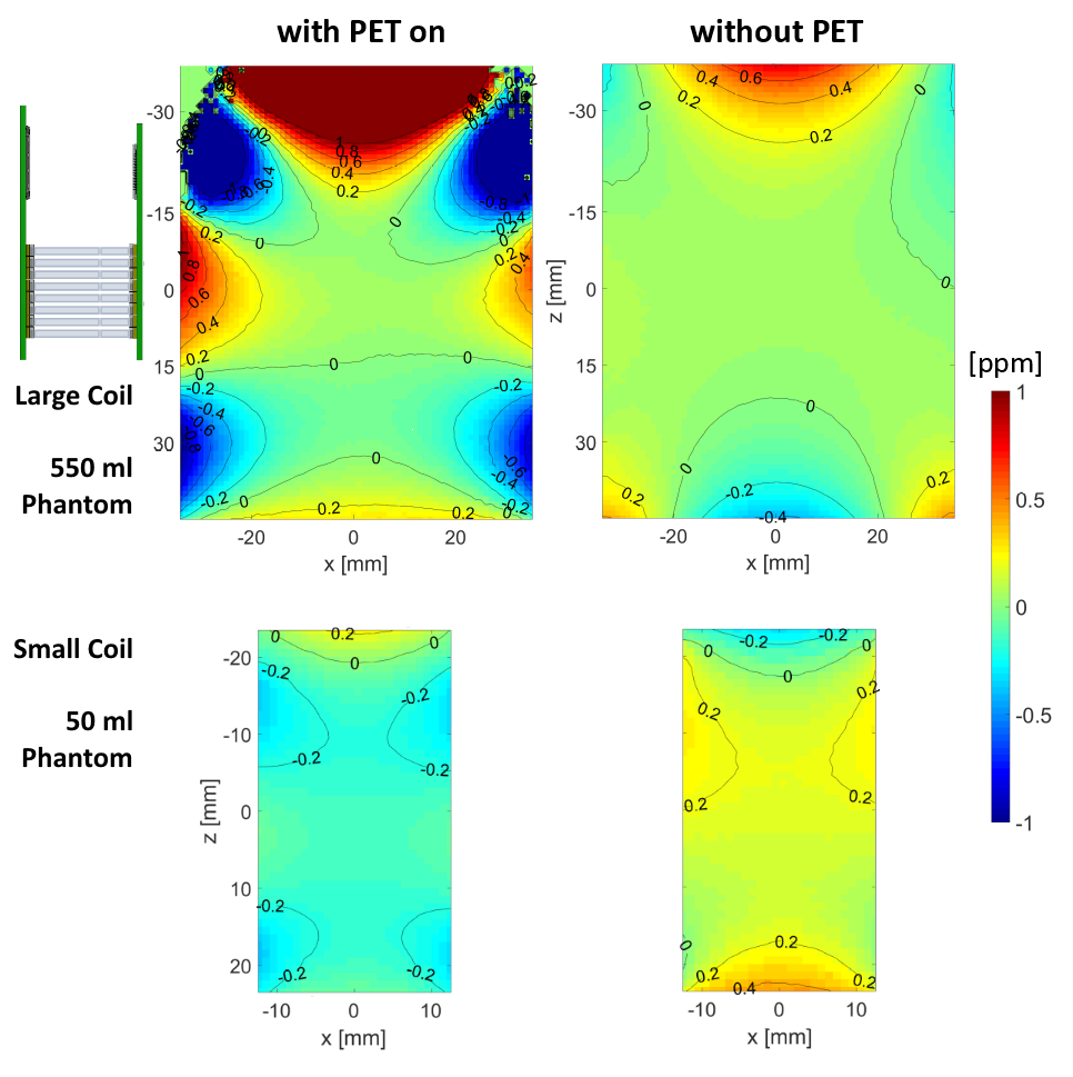
Horizontal slices from B0 static field maps, with and without the PET insert within or around volume coils. Distortions of +/- 1 ppm are seen on the edges of the large phantom and coil, near the locations of PET insert SiPMs and PET cable connections (left). With the small phantom and coil, there are negligible B0 distortions, because the MRI field of view with that coil extends only +/- 16 mm from the RF coil and magnet bore centre, well away from the PET components.
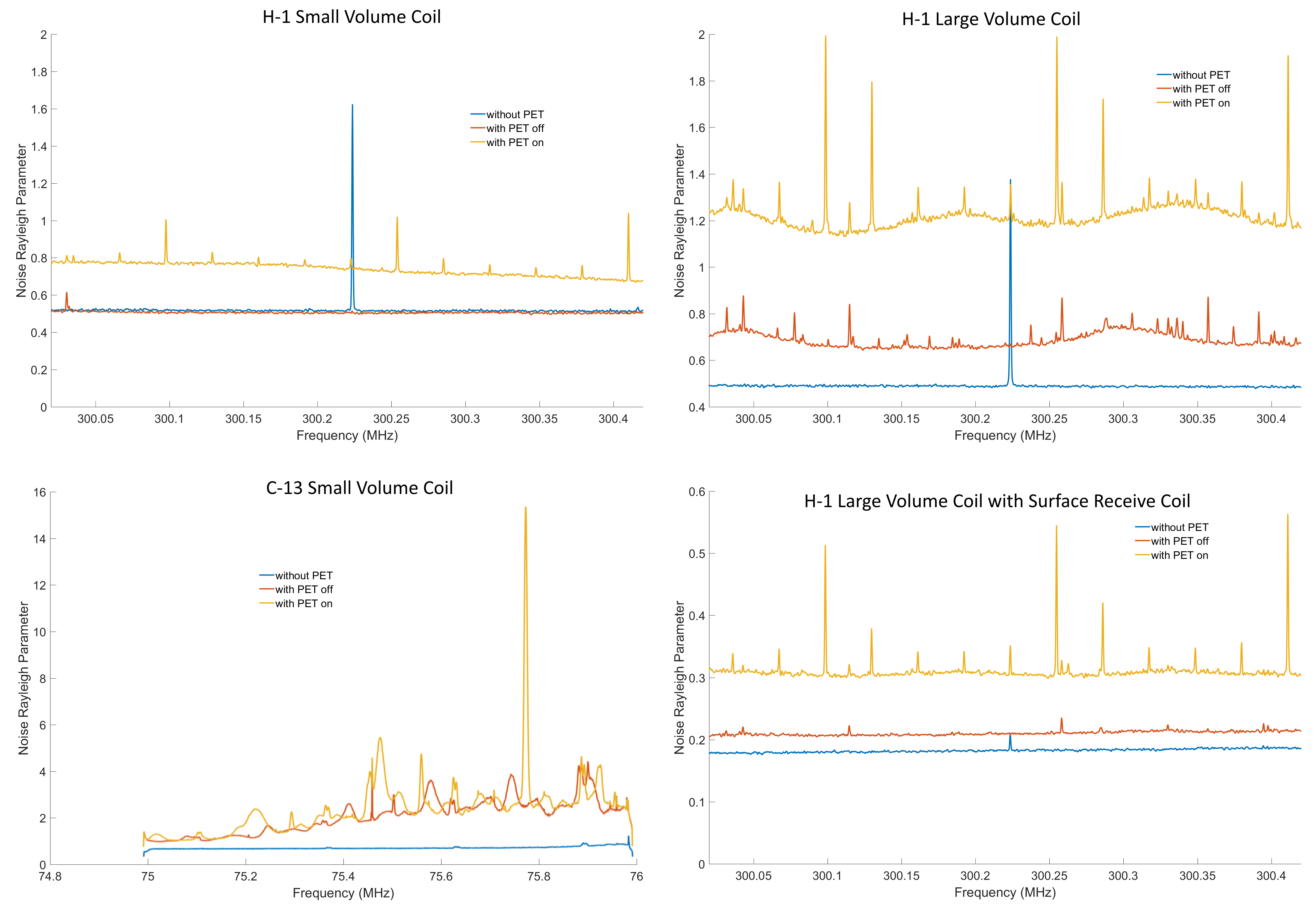
Noise spectra acquired, with various receive coil configurations, without insert, with inactive insert, and with acquiring PET insert. Data were acquired using a spectroscopy sequence with no gradients or RF transmit enabled. Signal magnitudes of 256 repetitions were fit with Rayleigh distributions for the Rayleigh width parameter, separately at each frequency bin.
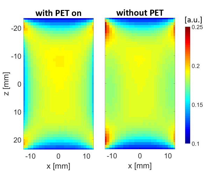
Horizontal B1 field maps, with and without the PET insert around the small volume coil, using a 50 mL uniform phantom filling the MRI RF volume coil bore. Gradient echo images were acquired with varying RF excitation voltage. At each pixel, signal was phase-corrected, and the rate at which real signal varied with voltage was fit for an effective B1 scale factor, which is plotted here in arbitrary units. Small differences are seen at the corners due to differences in the rotation of the volume coil; negligible differences appear due to the insert itself.
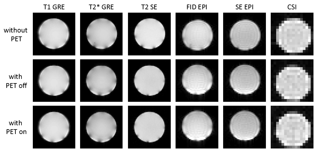
1H MRI and 13C CSI acquired of uniform phantoms without insert, with inactive insert, and with acquiring PET insert around the small volume coil.