1740
A comprehensive study on electrically floating PET insert for efficient RF penetrability at 3 T MRI system1National Institute of Radiological Sciences, Japan, Chiba, Japan, 2Stanford University, Stanford, CA, United States
Synopsis
A comprehensive experimental study has been conducted on the geometrical aspects of electrically floating radio frequency (RF) penetrable PET inserts to improve the RF penetration efficiency for acceptable MR imaging performance. Several one ring and two ring PET insert prototypes were used to do experiments in a 700-mm bore diameter 3 T clinical MRI system with a homogeneous cylindrical phantom. Study results provide guidance for optimized PET ring design for efficient RF field penetration inside the shielded ring.
Introduction
Locoregional PET inserts [1-4] for the existing MRI systems can become an affordable alternative to the commercially available body PET/MRI system. To avoid mutual interference between PET and MRI, PET detectors are shielded using conductive materials. An electrically floating PET insert [4] uses MRI built-in body RF coil as transmitter and/or receiver. The RF field from (and to) the body RF coil can pass through the narrow gaps in between the floating shielded PET modules – a grounded shield attenuates the field. However, because of large amount of shielding materials with the PET insert, the penetrated transmit RF field (B1) inside the PET ring is not strong enough to meet the basic requirement (e.g., required flip-angle) for practical MR imaging [4]. A comprehensive experimental study has been conducted in a 3T MRI system with prototype PET inserts to study on different geometric aspects of PET ring for improved RF penetration.Materials and Methods
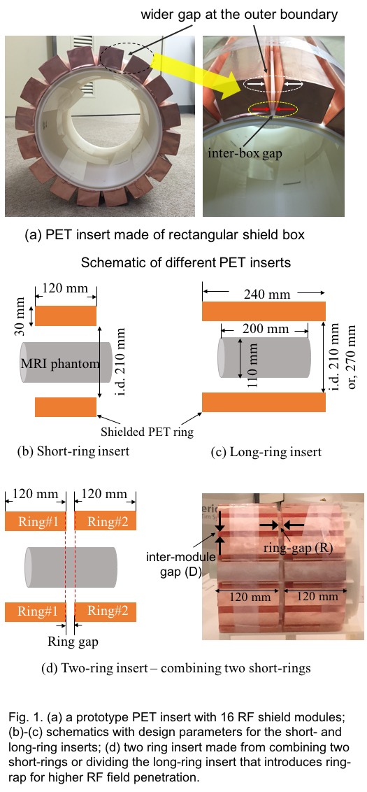
In this study, cylindrical PET ring with 16 empty RF shield boxes were used to study on the B1 field and SNR. Geometric parameters of the PET ring considered are: (a) axial length (240-mm vs. 120-mm); (b) inner diameter (210-mm vs 270-mm); (c) outer diameter (270-mm vs. 300-mm); (d) transaxial inter-module gap (1-mm vs. 3-mm); and, (e) axial gap (“ring-gap”) between two PET rings (5-mm, 10-mm and 20-mm). 21 shield boxes were used in case of larger inner diameter (270 mm) insert. Schematic representation with a photo of prototype two-ring PET insert are shown in Fig.1. All experiments were conducted in a 700-mm bore diameter 3T MRI system by using a 110-mm diameter and 200-mm axially-long cylindrical homogeneous phantom (NaCl and NiSO4 solution). The prototype insert with 240-mm and 120-mm axial lengths will be mentioned here as “long-ring” and “short-ring” insert, respectively. The two-ring insert was developed by combining two short-ring inserts with ring-gap in between. The shield boxes were made with 18 um thick copper foil.Results and discussion
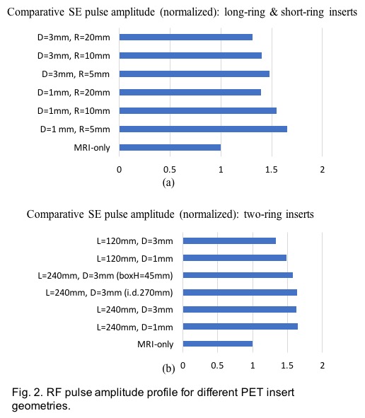
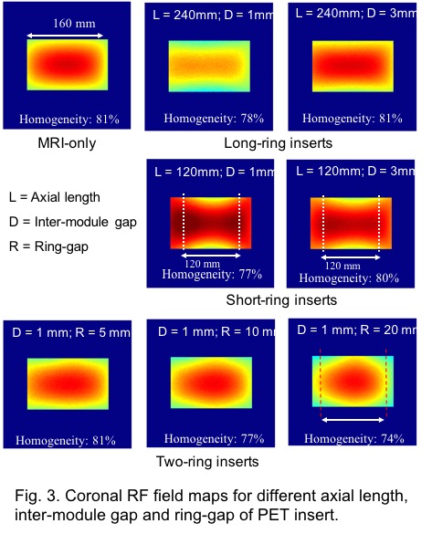
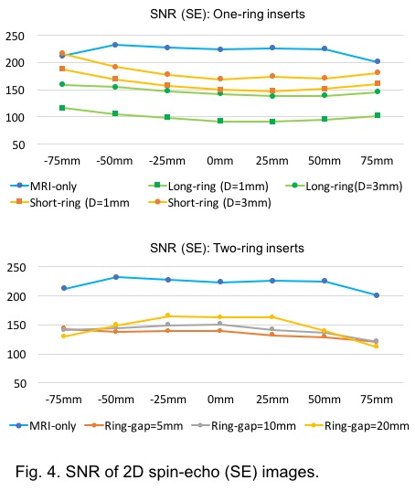
Some of the results are given here, in brief. Fig. 2 compares between long-ring, short-ring and two-ring inserts for the same coronal plane B1 map. It is evident that the long-ring insert exhibited high field attenuation when the inter-module gap was narrow (1-mm). On the other hand, for short ring insert high B1 field was found, since part of the phantom was outside the axial extent of the PET insert. From the RF pulse amplitude profile (Fig. 3), we found that, about 1.5 times more RF pulse amplitude were required for the PET inserts compared to the MRI-only case. This extra power is creating high RF field for the portion of the phantom that is outside the short-ring insert. On the other hand, in case of two-ring inserts (by dividing one long-ring into two short-ring insert), it was found that, even with narrow inter-module gap of 1-mm, the B1 field matched well with the MRI-only case which is mostly because of the RF field penetration through the ring-gap region. The comparative SNR results in Fig. 4 shows that short-ring exhibited higher SNR values compared to the long-ring. In case of two-ring insert, slight variation in SNR values was seen for the wider ring-gap inserts and mostly for imaging region near the ring-gap of the insert.
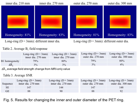
Study results for variation of inner and outer diameter of the PET ring (Fig. 5) did not show significant variation both in terms of B1 field and SNR values. Increasing inner diameter required higher number of shield boxes (from 16 to 21 for changing the inner diameter from 210-mm to 270-mm), which means number of inter-box gap increased by 5 compared to smaller inner diameter ring. For the 270-mm diameter ring, though the shielding materials increased, the increased number of inter-box gap compensated it. On the other hand, increasing the outer diameter (from 270-mm to 300-mm) for the same inner diameter (210-mm) provided wider outer diameter inter-module gap and that compensated the adverse effect of increased shielding materials for the large outer diameter ring by channeling higher RF field through the inter-box gap. For these reasons, we did not see much variation in RF pulse amplitudes as well.
Conclusion
Several conclusions can be made from the results given here: i) increasing inter-module gap improves the RF field penetration; ii) field penetration improves with the introduction of ring-gap for multiple-ring case even with reduced inter-module gap; iii) axial length of the PET ring should be carefully chosen; iv) increasing the inner dia. does not show much variation as the number of inter-module gaps increases as well; v) increasing the outer diameter also does not affect much.Acknowledgements
Part of this study was presented as poster to the ISMRM 2017 annual meeting.
References
[1] M. Schmand, et al, J. Nucl. Med., 48, 45P (2007). [2] K.J. Hong, et al, Med. Phys. 40, 1-11 (2013). [3] F. Nishikido, T. Obata, et al, Nucl. Instr. Meth. Phys. A, 756, 6–13 (2014). [4] P. Olcott, et al, Phys. Med. Biol., 60, 3459–478 (2015). [5] E. K. Insko, L. Bolinger, J. Magn. Reson. Series A, 103, 82-85 (1993). [6] L. Kaufman, et al, Radiology 173, 265-267 (1989).Figures