1730
Clinical Improvement of 19F Image Sensitivity using the Inductive Coupling at 7.0T Animal MRI11Division of Cellular and Gene Therapies, FDA/CBER/ Office of Tissues and Advanced Therapies, Silver Spring, MD, United States, 2Biomedical Physics, FDA/CDRH/Office of Science and Engineering Laboratories, Silver Spring, MD, United States
Synopsis
We present numerical simulations and experimental validation data testing the feasibility to improve 19F image sensitivity of perfluorocarbon labeled cells using the secondary resonator tuned at 287 MHz to make an enhancing induced RF magnetic field (B1) at 7.0T 19F/1H MRI. The numerical simulation results of |B1+| and corresponding experimental 19F images without and with the secondary resonator tuned at 287 MHz show the improvement of |B1+| and 19F image uniformity. To model a potential clinical application, we used inductive coupling MR to image 19F perfluorocarbon labeled cells encapsulated in polyethylene glycol (PEG) after their transplantation into mice.
Introduction
Commercially available transmit and receive surface coils often have simple planar designs that have a rapid fall-off in sensitivity with depth. Such limited radio frequency (RF) magnetic field (B1) uniformity is problematic when attempting to image even relatively small volumes of an animal anatomy. In our study, we demonstrate how the addition of a separate secondary resonator located beneath a commercial coil can provide uniform images of 19F nuclei with high sensitivity. Numerical simulations and experimental validation results show that the “secondary resonator” can be used to improve or worsen the image uniformity and sensitivity of 19F MRI.Methods
All experimental measurements were performed using an Agilent 7.0T horizontal bore animal MRI (Agilent Inc., Santa Clara, CA) with an open bore of 310 mm, a diameter of 115 mm inside the gradient coil (Resonance Research Inc., Billerica, MA). The commercial surface coil used for all experiments was a dual-tuned 19F/1H surface coil (RAPID MR International Inc., Columbus, OH). The secondary resonator (inner diameter (ID) = 18 mm, outer diameter (OD) = 22 mm) was tuned either to 287 MHz or 277 MHz with capacitors equal to 4.7, 5.5, and 11 pF (ATC Inc., Huntington Station, NY) combined with a variable capacitor to produce either an enhancing or an opposing RF magnetic field in the secondary resonator (B1L). A polyethylene glycol (PEG) disk with a diameter of 6 mm containing 12´106 19F labeled neural stem cells (NSCs) encapsulated in PEG following methods adapted from established protocols (1) was inserted subcutaneously in the back of a NSG mouse. The transplanted NSCs expressing luciferase were detected by bioluminescence after intraperitoneal injection of luciferin using the IVIS® SpectrumCT (PerkinElmer Inc., Waltham, MA). Numerical simulations were performed using the xFDTD software (Remcom Inc., State College, PA) and post-processing analysis was performed in Matlab (MathWorks Inc., Natick, MA). All the simulation results were normalized to obtain a magnitude of the transmit RF rotating magnetic field (|B1+|) equal to 2 mT at the center of the phantom.Results
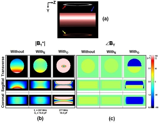
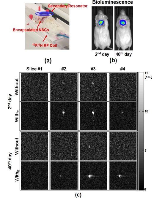
The |B1+| uniformity was improved when the resonator was tuned at 278 MHz (fL) with CL of 16.8 pF (Fig. 1, second column), whereas it becomes worse when fL was 277 MHz with CL of 18.0 pF (Fig. 1, third column). It shows that the enhancing induced B1+ would be produced when the secondary resonator was tuned at higher frequency compared to the resonance frequency of the 19F surface coil, fo of 282 MHz (Fig. 1, second column, WithE). Conversely, the opposing induced B1+ was produced when fL was lower than fo (Fig. 1, third column, WithO). Because the NSCs expressed the luciferase gene, the location and viability of the implanted cells could be monitored by luminescence over a period of 6 weeks (Fig. 2 (b)). MRI detection of the cells in vivo was done without (Fig. 2c, first row) and with (Fig. 2c, second row) the secondary resonator tuned at 287 MHz to make enhancing magnetic fields. The use of the resonator improved our ability to visualize transplanted cell location and survival non-invasively over a period of 6 weeks.Discussion
When comparing to the results available in literature related to our study of inductive coupling (2-3), our study presents the following novelties: 1) numerical simulation results of |B1+| and ÐBY (Fig. 1 (b) and (c)) without and with the secondary resonator designed for 19F imaging at 7.0T generated enhancing (second column) or opposing (third column) induced B1L depending on the tuning status of the resonator; 2) the method used in our study was based on in vitro and in vivo experimental results from a model clinical application, 19F labeled stem cells encapsulated in PEG (Fig. 2); 3) The in vivo MR imaging of encapsulated NSCs is a novel and potentially clinically relevant application of 19F MRI (Fig. 2). The use of a secondary resonator could be generally applicable in other similar situations where the available RF surface coil does not provide the desired image quality.
Disclaimer: “The mention of commercial products, their sources or their use in connection with material reported herein is not to be construed as either an actual or implied endorsement of such products by the Department of Health and Human Services.”
Acknowledgements
No acknowledgement found.References
1. Khetan S, Burdick J. J Vis Exp. 32, 1-5, 2009 2. Merkle H et al, MRM 66:901–910, 2011 3. Schnall et al., JMR 68:16 l-167, 1986Figures