1728
High Definition Sodium (23Na) In Vivo MRI of the Human Eye at 7.0 Tesla: Need for Substantially Enhanced Spatial Resolution than Commonly Used in Brain MRI1Berlin Ultrahigh Field Facility, Max Delbrueck Centrum, Berlin, Germany, 2MRI.TOOLS GmbH, Berlin, Germany, 3Institute of Radiology, Unviersity Hospital Erlangen, Erlangen, Germany, 4Division of Medical Physics in Radiology, German Research Centre (DKFZ), Heidelberg, Germany, 5Department of Ophthalmology, University of Rostock, Rostock, Germany, 6Institute of Physiology, Charite University Medicine, Berlin, Germany
Synopsis
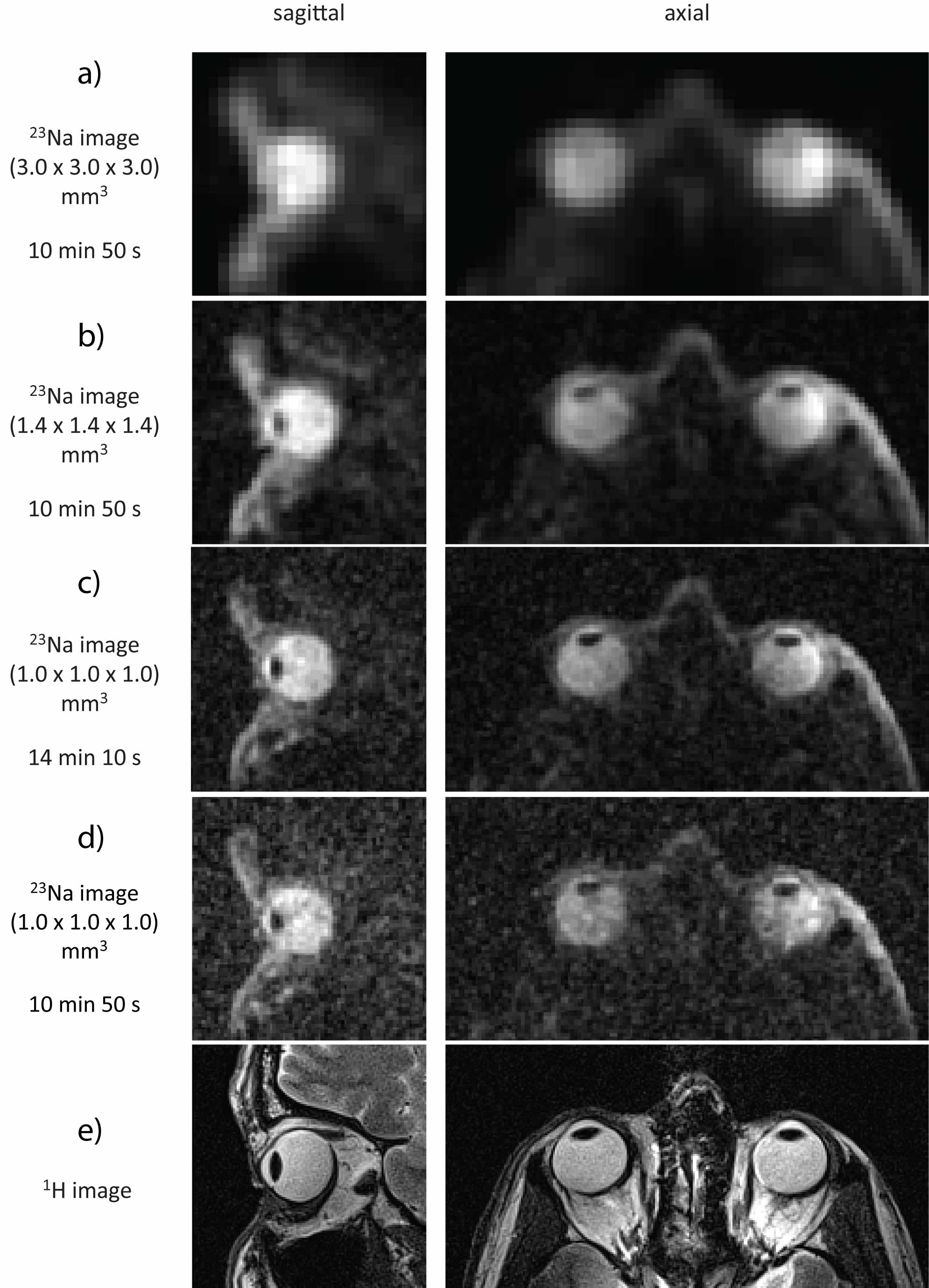
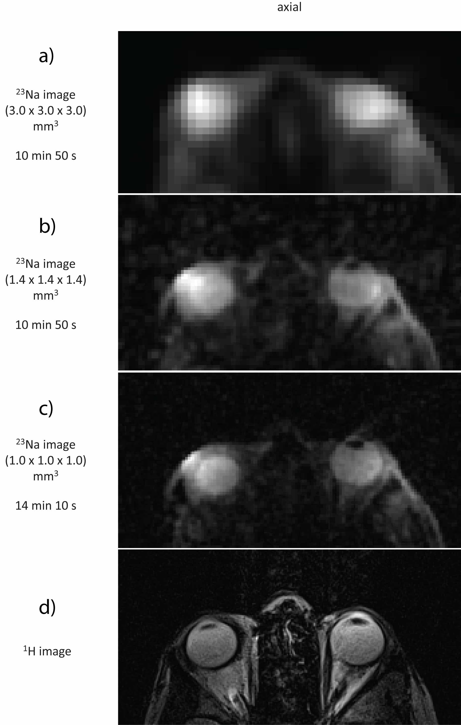
Sodium ions are crucial in the physiology of human eye and its compartments like vitreous humor, aqueous humor, lens and retina. In this work we used a six-channel transceiver array dedicated for ocular 23Na MRI and obtained in vivo images of the eye of exceptional quality with enhanced spatial resolution like (1.0x1.0x1.0) mm3 and demonstrated why spatial resolutions currently used for sodium MRI of the human brain are not sufficient in the context of 23Na in vivo MRI of the human eye. Enhancing spatial resolution is essential to investigate changes of sodium concentration in subtle eye compartments (aqueous humor, lens).
Introduction
Sodium ions (Na+) play a major role in the physiology of human organism and sodium/potassium pumps (Na+/K+-ATPase) are essential for the maintenance of homeostasis 1. Primary active transport, which is carried out by Na+/K+-ATPase, is undoubtedly a crucial link in processes which occur in the human eye and its compartments: the formation of the aqueous humor, the maintenance of sodium/potassium gradient between the lens and the vitreous humor, the removal of water and lactic acid from the retina 2. Once the function of sodium/potassium pump is impaired it might lead to disturbances in concentration/distribution of these ions what might be indicative of early pathological changes 3. 1H MRI demonstrated that imaging of subtle ocular structures requires a (sub)millimeter spatial resolution over a small field of view (FOV) 4. The goal of this study is to show why superior nominal spatial resolution (1.0 x 1.0 x 1.0) mm3 is absolutely essential for 23Na in vivo MRI of the human eye. Here we show in vivo sodium images of the human eye in order to benchmark the fidelity we obtained (1.0 x 1.0 x 1.0) mm3 with the proposed approach against nominal spatial resolution (3.0 x 3.0 x 3.0) mm3 which is typically used for 23Na MRI of the human brain 5.Methods
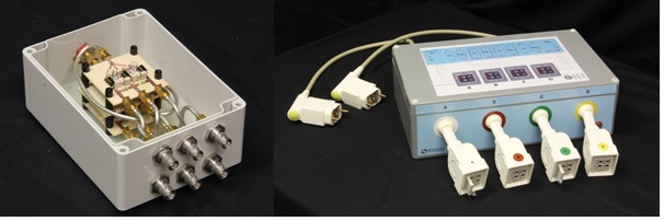
We used a six-channel transceiver array (Figure 1) which conforms very well to an average human head (Figure 1) along with a power divider which splits RF signal into six channels supporting equal amplitude and phase at all of the outputs (Figure 2). The multipurpose interface box (MRI.TOOLS GmbH, Berlin, Germany) (Figure 2) we used consists of 16 transmit/receive switches (Stark Contrasts, Erlangen, Germany): 8 for 1H and 8 for 23Na resonant frequency at 7.0 T along with integrated low-noise preamplifiers (Stark Contrasts, Erlangen, Germany). Human imaging studies were conducted on a 7.0 Tesla whole-body system (Magnetom, Siemens, Erlangen, Germany) using 3D-DAPR imaging technique for 23Na imaging and T2-weighted RARE imaging technique for proton imaging. Proton MRI was feasible by incorporating a single-tuned volume coil (Siemens, Erlangen, Germany).Results
We obtained sodium images from the eyes of two healthy, adult volunteers: one male (age = 53 years, BMI = 23.5 kg/m2) and one female (age = 28 years, BMI = 25.1 kg/m2) with nominal isotropic spatial resolution of (3.0 x 3.0 x 3.0) mm3, (1.4 x 1.4 x 1.4) mm3 and (1.0 x 1.0 x 1.0) mm3 (Figure 3a-c and Figure 4a-c). We also achieved an isotropic spatial resolution of (1.0 x 1.0 x 1.0) mm3 within 10:50 min scan time by reducing TR (Figure 3d). Images acquired with an isotropic spatial resolution of 3 mm are depicted in Fig. 3a (or Fig. 4a) and show a SNR which is superior to the high resolution datasets (Figure 3b-d and Figure 4b,c). Yet, all of the most important ocular compartments in the context of sodium physiology (vitreous humor, aqueous humor and lens) cannot be clearly delineated from the low resolution data. This shortcoming is resolved by using high spatial resolution 23Na MRI of the eye as highlighted in Figure 3c,d and Figure 4c. This finding underscores the need and value of high definition (isotropic spatial resolutions: ≤1mm) for 23Na in vivo MRI of the human eye.Discussion and Conclusion
Sodium in vivo MRI of the human eye using our methodology provides millimeter isotropic spatial resolution images of excellent quality obtained within clinically acceptable scan times. Our data demonstrate that applying higher spatial resolution (1.0 x 1.0 x 1.0) mm3 for 23Na eye imaging at 7.0 Tesla, what is made feasible by using a 6-channel transceiver array, clearly outperforms spatial resolutions currently used for brain imaging (3.0 x 3.0 x 3.0) mm3 in terms of revealed detail. The size of an average eye and – particularly - its substructures has major impact on the imaging protocols which will be used in future patient studies. For instance, mean axial and equator thickness of an average healthy human lens is about 4 mm. Mean anterior chamber depth, which is filled with aqueous humor, is around 3 mm. Typical intraocular tumors are between 1 mm and 3 mm in height and between 5 mm and 16 mm on the basis. Unlike a spatial resolution of 3 mm isotropic, a spatial resolution of 1 mm isotropic enables the delineation of subtle structures of the eye as well as potential pathologies.Acknowledgements
No acknowledgement found.References
1. Madelin G and Regatte RR, JMRI 2013
2. Kaufman PL, et al. Adler’s Physiology of the Eye, 2013
3. Wenz D, et al., ISMRM 2017
4. Graessl A, et al., Invest Radiol, 2014
5. Nagel AM, et al., Invest Radiol, 2011
Figures