1725
Optoelectronical-based multiplexed transmission of analog signals in a magnetic environment.1Aix-Marseille Univ, CNRS, CRMBM, Marseille, France, 2Aix-Marseille Univ, Polytech° Marseille, Ecole d'ingénieurs, Marseille, France, 3ESTIA Ecole supérieure des technologies industrielles avancées, Bidart, France, 4APHM, Hôpital Universitaire Timone, CEMEREM, Marseille, France
Synopsis
This study describes the methodological developments to both convert and transmit several mechanical signals in a magnetic environment (3T Verio Siemens) as optical signals. Multiple sensors were connected to a MR-compatible ergometer used to assess dynamic knee extensions kinetics. The corresponding signals were analog to digital converted and transmitted as optical signals through a single optical fiber. The quality of mechanical and 31P MR spectroscopy (31P-MRS) signals remained high and disclosed no adverse interference from the transducers ensuring both conversion and transmission. The multiplexed signals transmission allowed an accurate assessment of human movement kinetics in a magnetic environment.
Introduction
The assessment of activation and energetics in exercising muscle has been abundantly performed in high field magnetic environment1-3 using MR imaging (MRI) and 31P spectroscopy (31P-MRS). In order to control and measure exercise intensity, dedicated MR-compatible ergometers have been built4-6 and kinetic parameters of the movement have been assayed with specific sensors. For most of the setups reported so far, signals were transmitted with wires7,8 related to potential risks of electromagnetic interferences (EMI). More recently and with the aim of limiting EMI, a research group reported the utilization of an optical sensor9 which can be directly transmitted with an optical fiber where other used the conversion of analog signal from a conventional sensor to an optical transmission.5,10 However, these kinds of setup have never been designed for multiple sensors in order to assess multiple kinetic parameters leading to an accurate quantification of muscular work during exercise.
In the present study, we report results related to an original setup allowing a multiplexed optical transmission of signals from multiple analog sensors connected to MR-compatible ergometer. We demonstrated that multiple analog signals can be simultaneously transmitted via a single optical fiber.
Methods
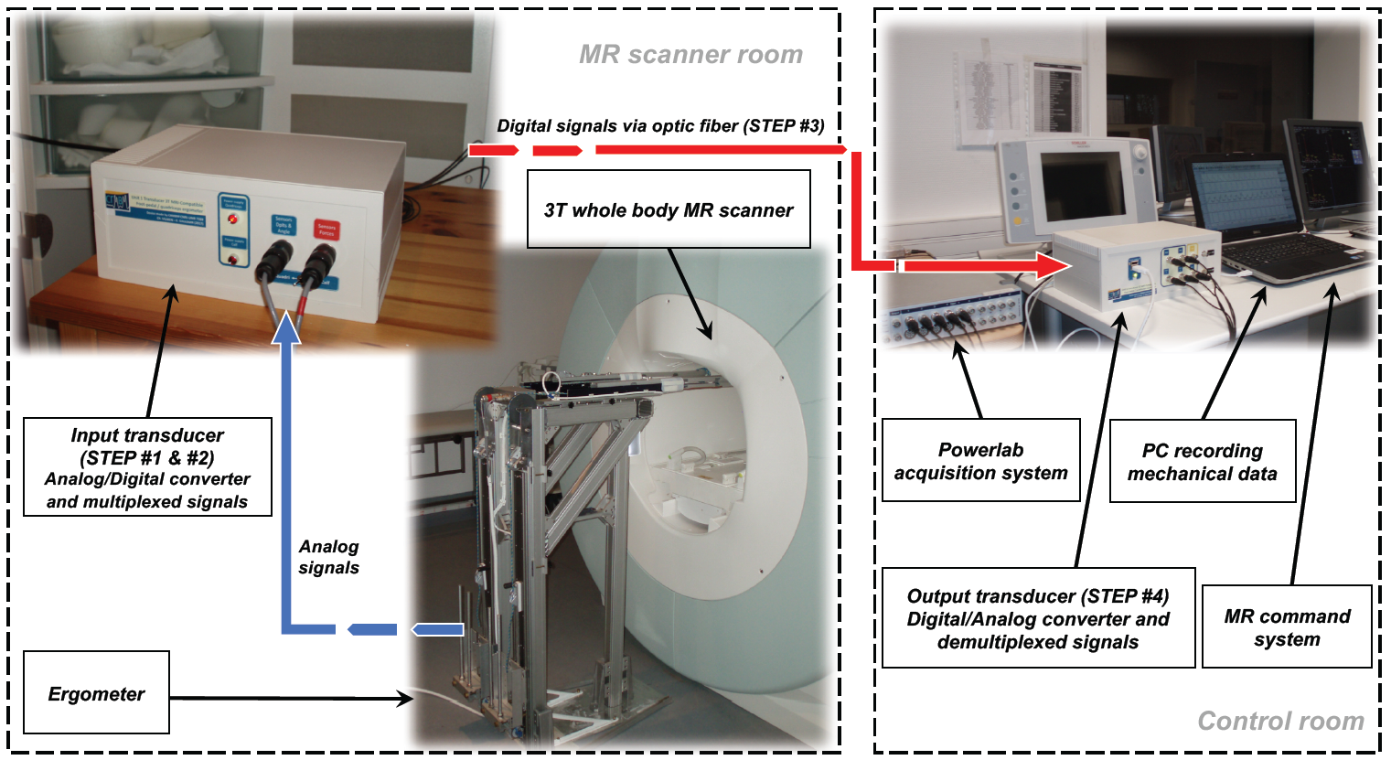
Two communication units connected by an optical fiber were designed to convert and transmit analog signals measured by sensors connected to a MR-compatible ergometer previously described.6 A schematic representation of the experimental design is displayed in Figure 1. Briefly, the unit in the MR scanner room digitally encodes the different analogic signals (forces, displacements and angle) and converts them into a multiplexed code using an Arduino-based microcontroller. Then the composite signal is transmitted (Steps #1 and #2) via a single optical fiber (Step #3) to the second unit located inside the control room. The second unit handles the composite signal demultiplexing and the digital-to-analog conversion, using another Arduino-based microcontroller (Steps #4).
In order to assess the accuracy of our measurements, we compared the analog signals at the sensors level and those from the output transducer. The corresponding results were analyzed with a Pearson product-moment correlation (r). A linear adjustment (calibration) between the electric signal and the calibrated measures was assessed with a determination coefficient R2.
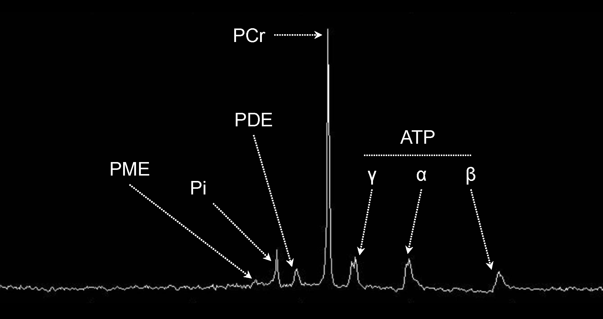
Analog signals were recorded during a 31P-MRS investigation performed in a single subject in a 3T super-conducting whole-body scanner (Siemens Verio, Erlangen, Germany). A 31P-1H surface coil was positioned under the right quadriceps femoris muscle group. Fully-relaxed (TR = 15s, NEX = 1, Number of Scans = 6) 31P spectra were recorded at rest while analog signals were measured during a submaximal exercise. The signal-to-noise (SNR) of 31P spectra and the PCr/ATP ratios were quantified.
Results
The correlations between analog signals measured on both sides of the optical transmission were very high (r > 0.99, Figure 2). The very high determination coefficients found for each channel (R² > 0.99) supported the accuracy of the corresponding measurements (Figure 2).
The SNR of the 31P MR spectra was very large (93 ± 9) while the averaged of PCr/ATP value was 3.9 ± 0.5 (Figure 4).
Discussion
We designed an optical-based electronic device which allowed accurate and reliable analog measurements in a magnetic environment. Shape and amplitude of mechanical signals were similar to those obtained in previous studies.6,11 The SNR of the 31P MR spectra and the metabolic ratio were also in accordance with those previously reported.12,13 In addition, our system considerably reduced wires congestion and potential electric/magnetic signal crosstalk. No mutual influence of the electric and magnetic signals was found thereby demonstrating the ability of our system to assess mechanical parameters in exercising muscle within a magnetic environment.Conclusion
Our electronic device based on a multiplexed transmission via an optical fiber allows an accurate conversion/transmission of several analog signals related to human movement kinetics and measured in a high-field magnetic environment.Acknowledgements
This study was supported by Centre National de la Recherche Scientifique (CNRS UMR 7339). The authors thank Dr. Sylviane Confort-Gouny for help in data acquisition and processing, the Assistance Publique des Hôpitaux de Marseille (APHM), and the subject who participated in the study.
References
1. Fouré A, Duhamel G, Vilmen C et al. Fast measurement of the quadriceps femoris muscle transverse relaxation time at high magnetic field using segmented echo-planar imaging. J Magn Reson Imaging. 2017;45:356-368.
2. Jubeau M, Y LEF, Duhamel G et al. Localized metabolic and t2 changes induced by voluntary and evoked contractions. Med Sci Sports Exerc. 2015;47:921-930.
3. Rossiter HB, Howe FA, Ward SA et al. Intersample fluctuations in phosphocreatine concentration determined by 31P-magnetic resonance spectroscopy and parameter estimation of metabolic responses to exercise in humans. J Physiol. 2000;528 Pt 2:359-369.
4. Quistorff B, Nielsen S, Thomsen C et al. A simple calf muscle ergometer for use in a standard whole-body MR scanner. Magn Reson Med. 1990;13:444-449.
5. Gusso S, Salvador C, Hofman P et al. Design and testing of an MRI-compatible cycle ergometer for non-invasive cardiac assessments during exercise. Biomed Eng Online. 2012;11:13.
6. Layec G, Bringard A, Vilmen C et al. Accurate work-rate measurements during in vivo MRS studies of exercising human quadriceps. MAGMA. 2008;21:227-235.
7. Tschiesche K, Rothamel M, Rzanny R et al. MR-compatible pedal ergometer for reproducible exercising of the human calf muscle. Med Eng Phys. 2014;36:933-937.
8. Liu JZ, Dai TH, Elster TH et al. Simultaneous measurement of human joint force, surface electromyograms, and functional MRI-measured brain activation. J Neurosci Methods. 2000;101:49-57.
9. Sinha S, Shin DD, Hodgson JA et al. Computer-controlled, MR-compatible foot-pedal device to study dynamics of the muscle tendon complex under isometric, concentric, and eccentric contractions. J Magn Reson Imaging. 2012;36:498-504.
10. Jeneson JA, Schmitz JP, Hilbers PA et al. An MR-compatible bicycle ergometer for in-magnet whole-body human exercise testing. Magn Reson Med. 2010;63:257-261.
11. Foure A, Wegrzyk J, Le Fur Y et al. Impaired mitochondrial function and reduced energy cost as a result of muscle damage. Med Sci Sports Exerc. 2015;47:1135-1144.
12. Fiedler GB, Schmid AI, Laistler E et al. Signal-to-noise ratio analysis of ³¹P MRS in skeletal muscle: influence of localization schemes, field strength, RF coils and channel phasing. 30th Annual Scientific Meeting of the European Society for Magnetic Resonance in Medicine and Biology 2013.
13. Rico-Sanz J, Zehnder M, Buchli R et al. Noninvasive measurement of muscle high-energy phosphates and glycogen concentrations in elite soccer players by 31P- and 13C-MRS. Med Sci Sports Exerc. 1999;31:1580-1586.Figures
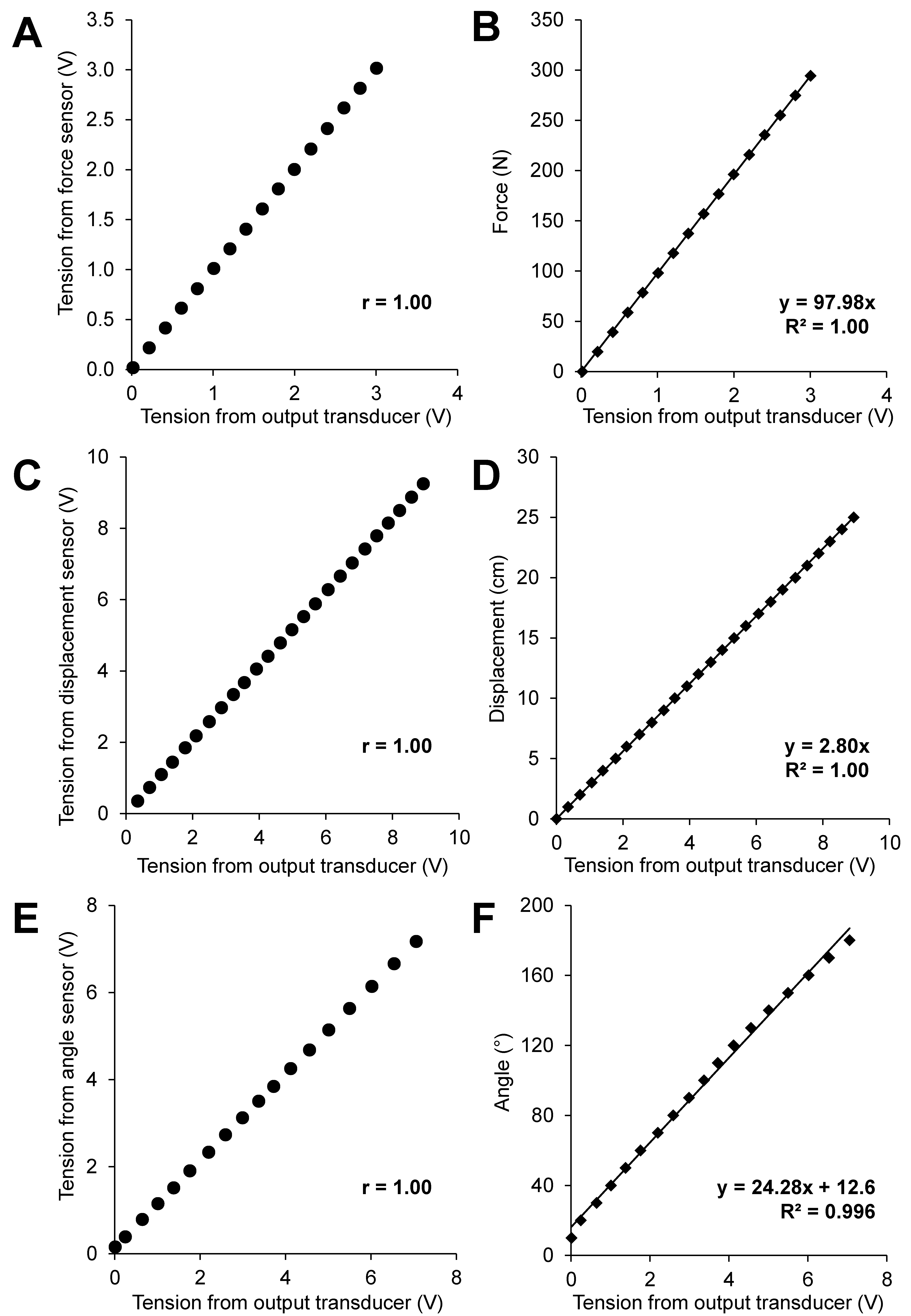
Correlations between analog signals coming out from the MR compatible ergometer and analog signals received after the optical-based transmission (A, C and E). Linear adjustments of output signals and calibrated measures (B, D and F) for three different mechanical signals including knee extension force (A and B), foot displacement (C and D) and knee angle (E and F).
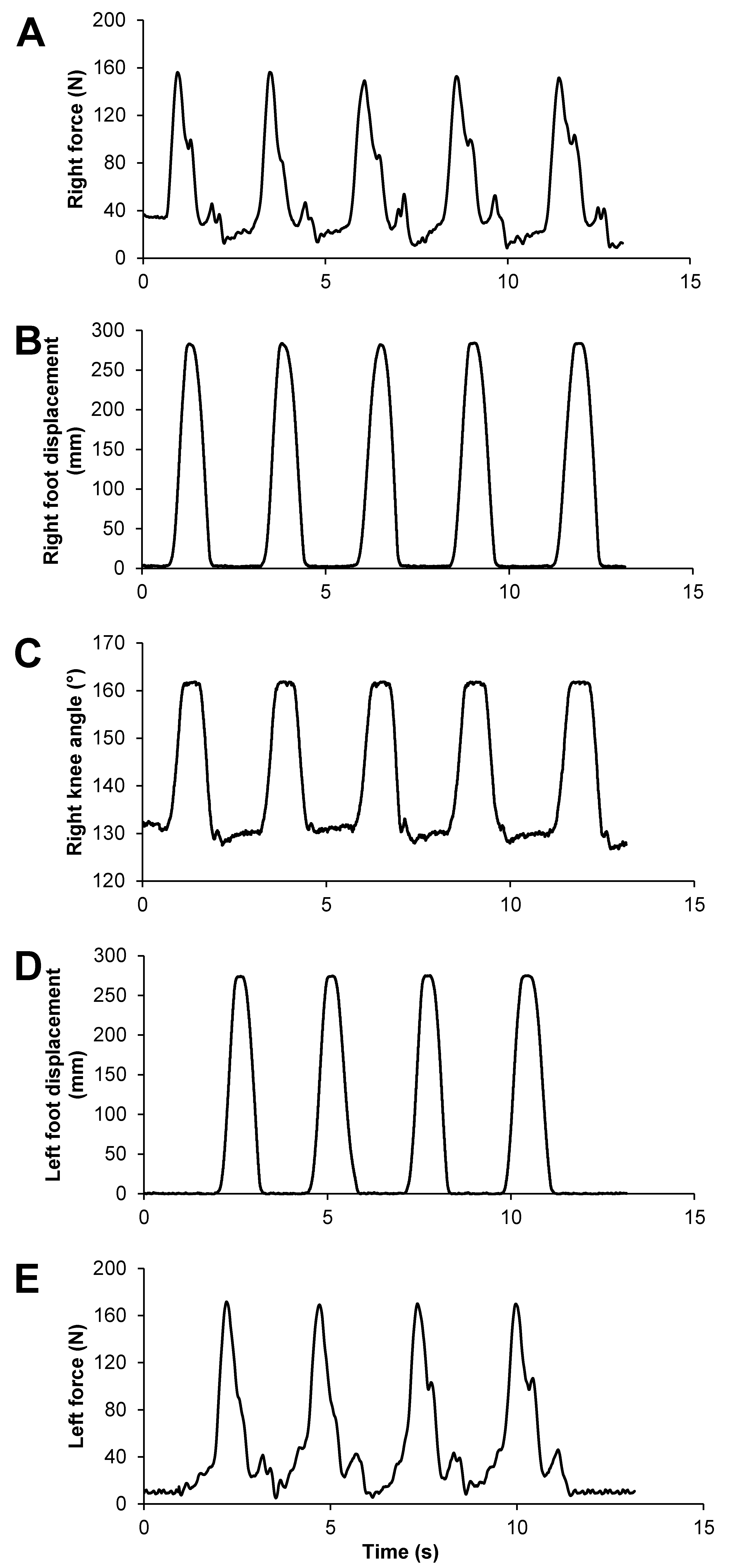
Typical raw mechanical signals including knee extensor force (A and E), foot displacement (B and D) and right knee angle (C) obtained during a submaximal voluntary concentric exercise.