1717
A genuine design for a dual-tuned $$$^{1}H/^{31}P$$$ coil with no lumped elements operating at 4.7T1Department of Nanophotonics and Metamaterials, ITMO University, Saint-Petersburg, Russian Federation, 2CNRS/CRMBM, Aix-Marseille University, Marseille, France, 3CNRS/Institute Fresnel, Aix-Marseille University, Marseille, France
Synopsis
For a wide range of MRI and MRS applications dual-tuned MR coils are used capable of multi-nuclear studies. Conventional ultra-high-field preclinical dual-tuned coils are either surface loops having high SNR over a limited FOV or volumetric coils with ultimate coverage compromised by low SNR while used in Tx and Rx regimes. In this contribution we propose an alternative design of the dual-tuned 1H/31P coil based on an open self-resonant periodic structure, which doesn’t require variable lumped capacitors for tuning and matching. It has been shown that the proposed coil is suitable for studying energetics in human forearm muscles at 4.7T.
Purpose
Dual-tuned coils are of a great interest in MRI and MRS due to their applicability for multi-nuclei studies. Conventional dual-nuclei coils for preclinical applications are basically surface loops or volumetric coils (e.g. birdcages) tuned and matched at the desired frequencies by means of at least four non-magnetic capacitors [1,2]. Surface loops introduce high intrinsic SNR but for limited ROIs due to the strong localization of the RF magnetic field in the proximity of the coil. In the opposite way, birdcages have ultimate coverage of large areas of interest but with a low SNR over the whole FOV when used in both Tx and Rx regimes. In this work we propose an alternative multi-mode resonator and a new corresponding dual-tuned 1H/31P coil, which we designed and experimentally demonstrated for studying energetics in human forearm muscles at 4.7T.Methods
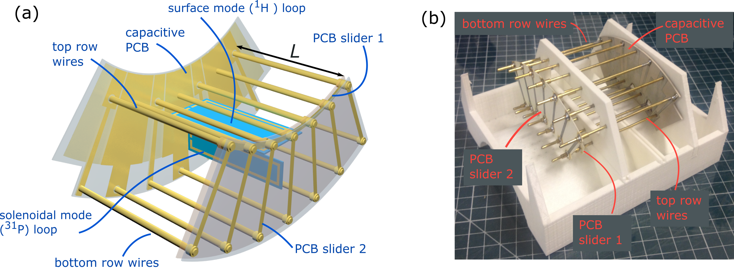
The coil comprised a parallel-wire resonator based on an open self-resonant periodic structure, consisted of two rows of seven parallel subwavelength wires each (Figure 1(a)) [3,4]. Two mutually orthogonal eigenmodes of the resonator were excited through inductive coupling by the two small magnetic loops: the surface mode at 200 MHz (1H) and the volumetric (solenoidal) mode at 82 MHz (31P). To tune the resonator for a wide range of different loadings without variable capacitors at the two Larmor frequencies, two sliding connectors were attached to the wires. The first one (PCB Slider 1) connects all the top row wires affecting the surface mode only, while the second one (PCB Slider 2) shortcuts each top-bottom wire pairs and tune the volumetric mode by sliding. At the opposite end of the parallel-wire structure the wires were paired by interconnecting with two different values of capacitance, distributed on a common PCB. Matching at both frequencies was provided by varying the inductive coupling between the resonator and both the feeding loops, achieved by adjusting of the distances to the latter.
The proposed coil was simulated in Frequency Domain Solver of CST Studio. Two rows of 7 wires were represented by 2mm diameter brass tubes. The feeding loops as well as constructive capacity and the sliders were implemented as thin copper strips on Arlon AD1000 0.5-mm-thick substrate. The model also contained the 130mm-diameter RF-shield of a 4.7T MR scanner and a homogeneous cylindrical phantom (radius 50mm, length 250mm, ε=34, σ=0.4 S/m).
The proposed coil was manufactured and tested on the bench and in the environment of an MR scanner. Both feeding loops were connected through separate 50-Ohm coaxial cables to the MR system. The resonator was inserted to a 3D-printed holder conformal to the shape of the human arm (Figure 1(b)). Simultaneous operation of the coil at 1H and 31P frequencies was tested by measuring the S-parameters of the two coaxial ports with the vector network analyzer Anritsu MS2036C. MRI and MRS application of the proposed coil was shown on the Bruker BioSpec 4.7 Tesla scanner: a homogeneous 0.9% saline water phantom containing a phosphorus compound was scanned using FLASH sequence (TR/TE=100/4.3ms, voxel size 0.57x0.57x5mm). 31P non-localized spectra of the phantom were also obtained by means of a single pulse-acquire sequence (TR=1500ms, NS=16).
Results
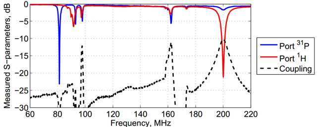
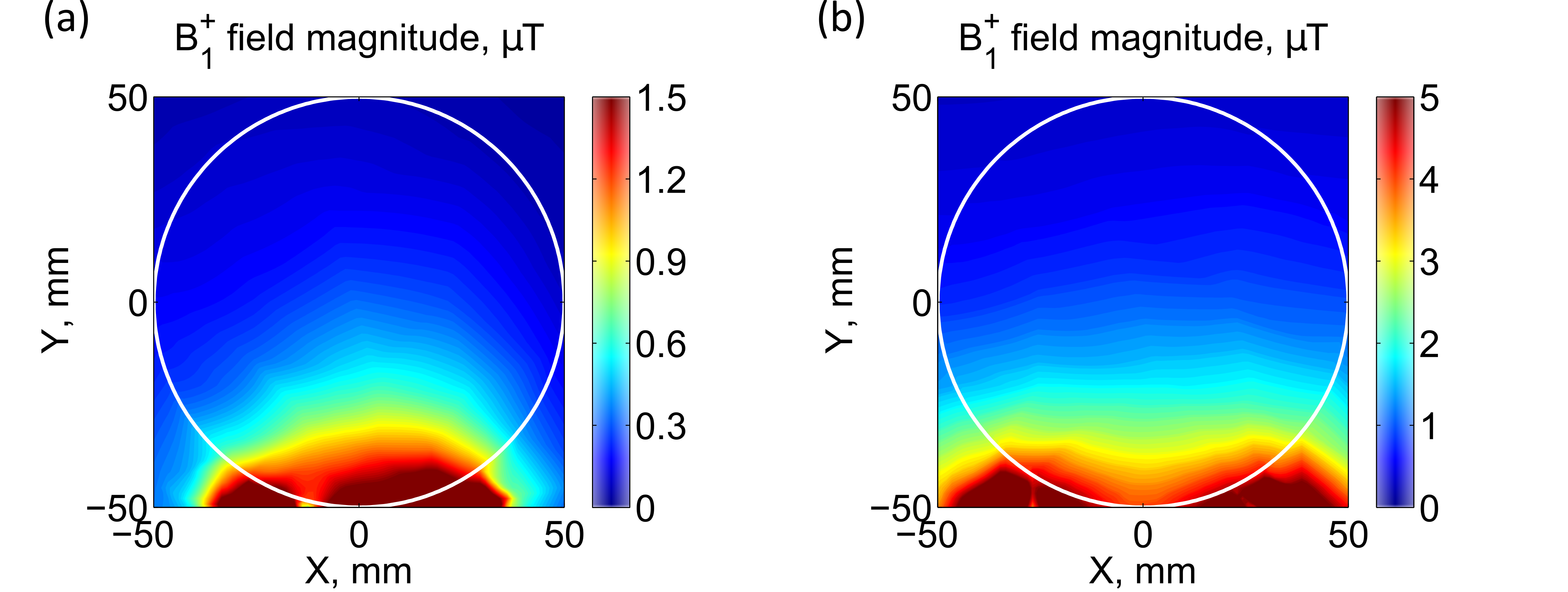
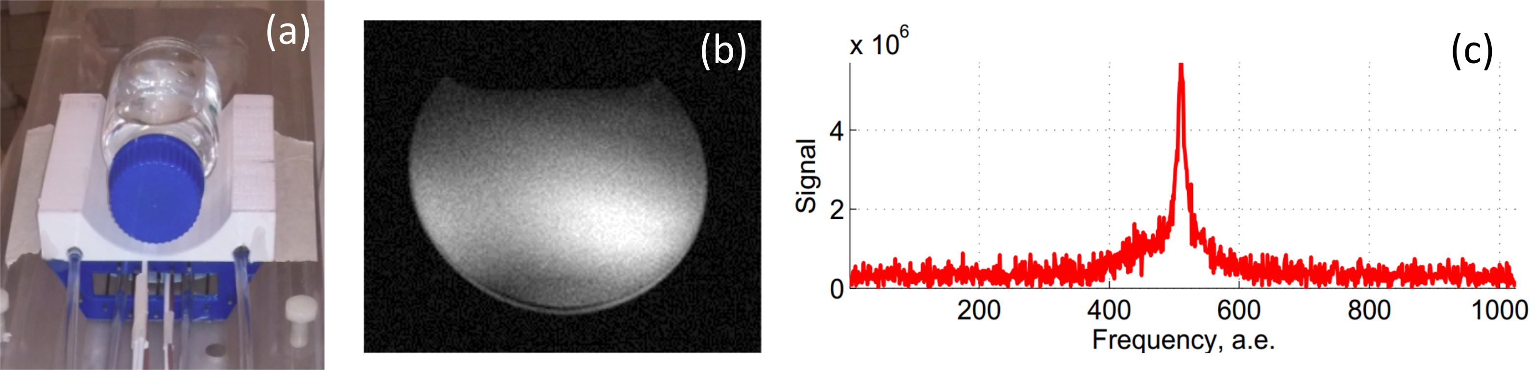
Figure 2 illustrates the behavior of measured S-parameters of the proposed coil in the 60 – 220MHz frequency range. It shows tuning and matching of 31P and 1H ports (reflection coefficient magnitude below -20 dB both at 82 and 200 MHz) as well as high isolation between the ports (coupling coefficient below -11dB). Simulated B1+ maps of the coil for both 1H (a) and 31P (b) frequencies are depicted in Figure 3. The magnetic RF field at both frequencies is confined within the top muscle region, which gives a desired spatial selectivity in spectroscopy tasks. For a bottle phantom, positioned on the top of the coil (Figure 4(a)), the obtained 1H axial-plane image is shown in Figure 4(b) and the corresponding non-localized 31P spectrum, obtained without retuning of the coil, is shown in Figure 4(c).Conclusion
We designed a novel dual-nuclei (1H/31P) MR coil for muscle energetics MRI/MRS applications at 4.7 Tesla. The proposed coil design is based on a parallel-wire self-resonant periodic structure with structural capacitance. The coil satisfied tuning and matching conditions at both the 1H and 31P Larmor frequencies thanks to mechanically adjustable positions of the tuning sliders and the feeding loops with no lumped capacitors required. Moreover, we have shown preliminary results of MR tests of the coil in 1H imaging and 31P non-localized spectroscopy on a phantom. The results confirm the proper field of view desired for the selected application.Acknowledgements
This work was supported by the Ministry of Education and Science of the Russian Federation (project No. 14.587.21.0041 with the unique identifier RFMEFI58717X0041).
This project has received funding from the European Union's Horizon 2020 research and innovation program under grant agreement No 736937.
Acknowledgments to Institute Carnot Star, meta twins project.
References
[1] Alecci M. et al., “Practical design of a 4 Tesla double-tuned RF surface coil for interleaved 1H and 23Na MRI of rat brain”, Journ. of Magn. Reson., vol. 181(2), p. 203–211, 2006
[2] J. Thomas Vaughan, John R. Griffiths, “RF Coils for MRI”, John Wiley and Sons Ltd, 2012.
[3] Slobozhanyuk A.P. et al., “Enhancement of Magnetic Resonance Imaging with Metasurfaces”, Adv. Mater., vol. 28, p. 1832–1838, 2016
[4] Jouvaud C. et al., “Volume coil based on hybridized resonators for magnetic resonance imaging”, Appl. Phys. Lett., vol. 108(2), p. 023503, 2016
Figures