1709
A double resonant (1H/23Na) whole-body RF system for MRI at 3TMatthias Malzacher1, Nadia Paschke1, Jorge Chacon-Caldera1, and Lothar R. Schad1
1Computer Assisted Clinical Medicine,Medical Faculty Mannheim, Heidelberg University, Mannheim, Germany
Synopsis
23Na MRI keeps increasingly demonstrating diagnostic value in a multitude of studies and clinical applications due to its capability to provide information on tissue viability. In order to co-register 23Na and 1H MR images, a double resonant 23Na/1H RF system is the optimal solution. In this work we present a clinical double-resonant RF system consisting of a shielded 23Na BC coil, a 16 channel 23Na Rx array and a local 1H Helmholtz coil inside the shielded 23Na BC coil. The complete system is demonstrated in EM simulations and initial feasibility measurements are performed.
Introduction
23Na-sodium MRI keeps increasingly demonstrating diagnostic value in a multitude of studies and clinical applications due to its capability to provide information on tissue viability[1,2]. Yet, sodium MRI suffers from weak SNR and low abundance of sodium in the human body.Therefore, 1H MR images are needed for morphological information to compensate for the poor resolution of sodium MRI. In order to co-register 23Na and 1H MR images, a double resonant 23Na/1H RF system is the optimal solution. Since clinical scanners do not support X-nuclei hardware integrated in the MR system, local coils have to enable combined proton and X-nuclei MRI. In this work we present a clinical double-resonant RF system consisting of a shielded 23Na BC coil[3], a 16 channel 23Na Rx array and a local 1H Helmholtz coil inside the shielded 23Na BC coil. The complete system is demonstrated in EM simulations and initial feasibility measurements are performed.Methods
Simulation:EM fields were calculated using FEM simulations (CST – Computer Simulation Technology GmbH) with a tetrahedral mesh. Mesh refinement converged for all simulations to at least 5% for all S-parameters at the resonance frequency 32.586 MHz (23Na) and 123.2 MHz (1H).
23Na Tx:
A quadrature driven 16-legged asymmetric 23Na was modeled following reference[3].
23Na Rx:
A 16 channel 23Na Rx only array was designed with 8 posterior and 8 anterior channels to be used for the abdomen. The Rx elements were decoupled iteratively using EM simulations. Each Rx coil was split twice and a 1H trap was added. The H-fields of the 23Na Rx channels were combined using the matched filter approach[4].
1H TxRx:
The 1H TxRx coil was constructed as a Helmholtz coil covering the FoV of the 23Na Rx array. Each loop of the Helmholtz coil was equally split 16 times.
Phantom:
Four cylindrical phantoms were used with diameter = 115 mm, length = 205 mm, σ = 0.91 S/m and ε = 80.
The simulation setup of the different coils separately and the complete simulation setup are shown in Figure 1 a).
Measurement:
For initial feasibility measurements, the posterior part of the setup consisting of an 8 channel 23Na Rx only coil (Figure 2) combined with one 1H TxRx posterior coil was built using the dimensions of the EM simulation. Four cylindrical phantoms (Siemens Healthineers, dimensions and solution as described above) were used for phantom measurements.
We used a density adapted 3D radial sequence[5] for sodium imaging (TE/TR/FA=0.54ms/50ms/54°, 8500 projections, FoV=(450mm)3, resolution=(6mm)3, TRO=20ms), and a 2D gradient echo sequence for proton imaging (TE/TR/FA=10ms/358ms/90°, FoV=450mm², matrix=256²,Res = 1.75 mm, slice=5mm).
The reference scan was acquired using the 23Na BC coil stand alone in TxRx mode.
In order to protect the detuning PIN diodes of the 23Na BC coil it stayed resonant during the 1H measurement but the 23Na Rx array was detuned.
For SNR calculation a noise scan was performed using the same parameters as for the signal scan but without Tx power applied. The 23Na Rx channels were combined identical to the simulation using the matched filter approach.
Results
Simulation:The center coronal planes of the simulated B1-maps (normalized to 1W accepted power) are shown in Figure 1 b). The 23Na array showed a 2-fold B1-field increase compared to the 23Na BC coil. The 23Na array yielded an up to 6-fold higher B1-field compared to the 23Na BC coil near the Rx coils.
The 1H TxRx coil covered almost the whole FoV of the 23Na Rx array but showed a slightly unbalanced behavior in the longitudinal direction.
Measurement:
The center coronal planes of the SNR measurements are shown in Figure 3. Consistent with the simulations, the 8 channel 23Na Rx only array already achieved a 2-fold SNR increase in the coronal plane and an up to 6-fold SNR increase near the Rx coils compared to the 23Na BC coil.
The 1H SNR map revealed a similar FoV as the 23Na Rx only array. Yet, the coil profile was unbalanced in longitudinal direction.
Discussion & Conclusion
A double resonant 1H/23Na whole-body RF setup was demonstrated for clinical MRI at 3T. The feasibility of a 16 channel 23Na Rx only array combined with a 1H Helmholtz coil inside a 23Na BC coil was proven in simulation. A 2-6 fold improvement of the 23Na array was achieved over the whole FoV in simulation. This performance could also be reproduced in initial SNR measurements of a reduced setup. Yet, the rudimentary 1H coil revealed an unbalanced coil profile and low SNR. Future research should focus on getting 1H performance near to a clinical level. The RF setup should additionally be evaluated in in-vivo scans.Acknowledgements
No acknowledgement found.References
[1] Madelin G, Ravinder RR. Biomedical applications of sodium MRI in vivo. J Magn Reson Im. 2013;38(3):511-529.[2] Zöllner FG, Konstandin S, Lommen J, et al. Quantitative sodium MRI of kidney. NMR Biomed. 2016;29(2):197-205.
[3] Wetterling, F., Corteville, D.M., Kalayciyan, R. et al., Whole body sodium MRI at 3T using an asymmetric birdcage resonator and short echo time sequence: first images of a male volunteer. Phys Med Biol, 2012;57(14), p.4555.
[4] Schnell W, Renz W, Vester M, Ermert H. Ultimate signal-to-noise-ratio of surface and body antennas for magnetic resonance imaging. IEEE T Antenn Propag, 2000;48(3):418-28.
[5] Nagel AM, Laun FB, Weber MA, et al. Sodium MRI using a density‐adapted 3D radial acquisition technique. Magn Reson Med.2009;62(6):1565-1573.
Figures
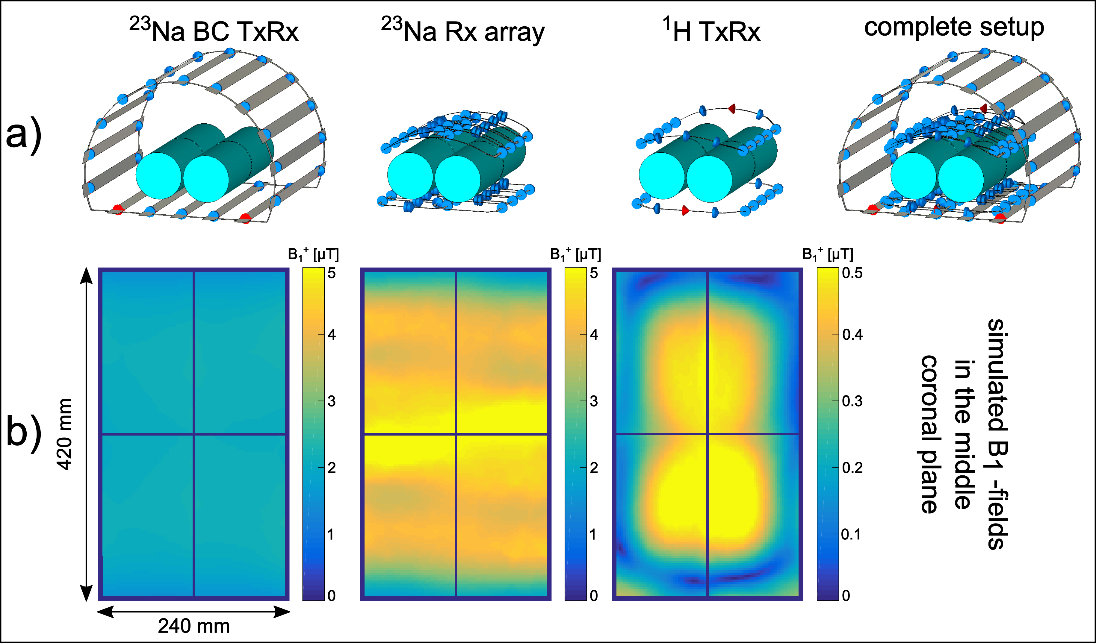
a) Simulation setups of the different coils separately and
the complete setup combined including the bottle shaped phantoms. b) Simulated
B1-field (normalized
to 1W accepted power) in the center coronal plane. The H-fields of the 23Rx
channels were combined using the matched filter approach[4].

23Na 8 channel Rx array
including preamplifier, detuning circuits and 1H trap circuits.
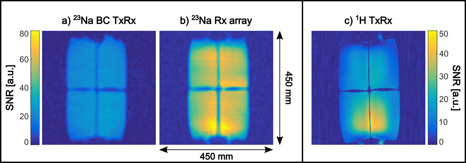
Measurement
results of a) the 23Na BC coil in TxRx mode, b) the 23Na Rx only array and c)
the 1H TxRx coil.