1675
Prediction of histological grade of hepatocellular carcinoma using quantitative diffusion-weighted magnetic resonance imaging: a retrospective multi-vendor study1Tokyo Medical and Dental University, Tokyo, Japan, 2JA Toride Medical Center, Ibaraki, Japan
Synopsis
Eighty-three patients with 100 histologically diagnosed hepatocellular carcinomas (HCCs) who preoperatively underwent diffusion-weighted (DW) imaging at any of 6 institutes were retrospectively studied. Receiver-operating characteristic analysis revealed that quantitative measurements such as the relative contrast ratio (RCR) and the contrast-to-noise ratio (CNR) between lesion and liver parenchyma on DW images were superior to the apparent diffusion coefficient (ADC) in predicting poorly differentiated HCCs, and intraclass correlation coefficients for the RCR tended to be greater than for the CNR and the ADC.
Introduction
Histological grade is one of the prognostic factors in patients with hepatocellular carcinoma (HCC)1-4. Accurate radiological stratification of HCC grade is helpful for predicting the prognosis and selecting the therapeutic strategy.
The apparent diffusion coefficient (ADC), which is the most commonly used index in diffusion-weighted MR imaging (DWI), has been used for quantitative assessment of malignant tumors. Several studies have evaluated the correlation between the ADC and histological grade of HCC5-14 but yielded conflicting results. Recently, some studies showed that non-ADC parameters, i.e., the relative contrast ratio (RCR) or contrast-to-noise ratio (CNR) of HCC to the surrounding liver parenchyma on DW images, were superior to the ADC in predicting the grade of HCC.6, 7, 9 However, they were single-center studies using single magnetic resonance (MR) units, and the inter-observer reproducibility of these measurements has not been reported.
The aim of this study was to evaluate the usefulness and the reproducibility of DW parameters, including RCR, CNR and ADC using multi-vendor MR units for predicting grade of HCC.
Methods
This multi-center, retrospective, multi-vendor study included patient data from 6 institutions. The study protocol was approved by the institutional review boards at 3 institutions. The other 3 Institutions did not have ethical review committees so delegated approvals to the main institution. The requirement for informed consent was waived.
Eighty-three patients with 100 histologically diagnosed HCCs who underwent preoperative liver DW imaging with b = 0 and 1000 s/mm2 at any of 4 institutions (49 patients with 58 HCCs) or b = 0 and 800 s/mm2 at any of 2 institutions (34 patients with 42 HCCs) were included. Two radiologists independently measured the ADC of the lesion as well as the RCR and the CNR between the lesion and the liver parenchyma on high b-value DW images. The diagnostic performance of the DW parameters in discriminating poorly differentiated HCCs was compared using receiver-operating characteristic (ROC) analysis. Inter-observer agreement between the radiologists was assessed using the intraclass correlation coefficient (ICC).
Results
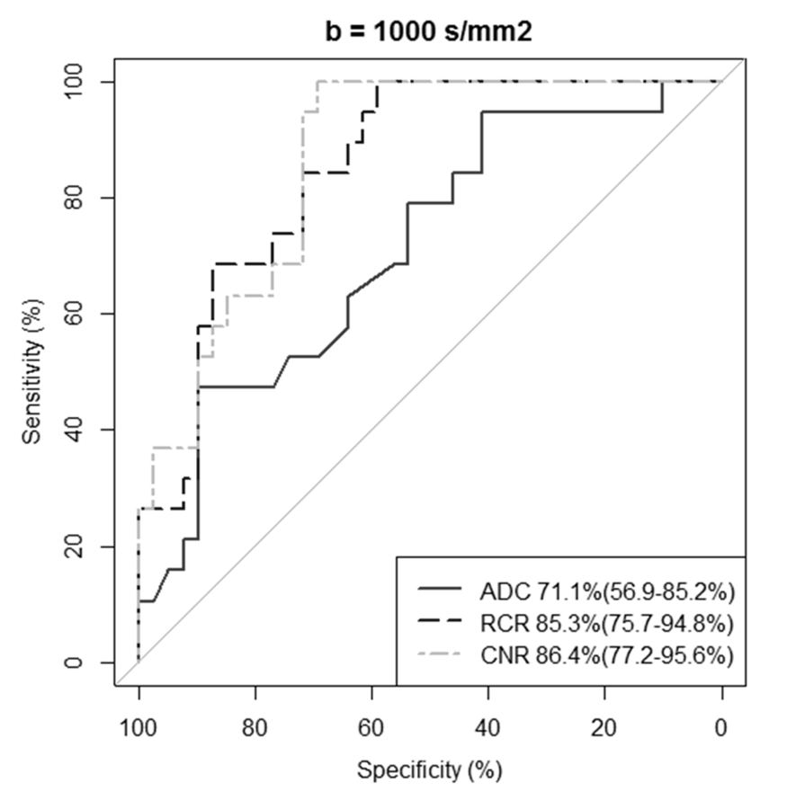
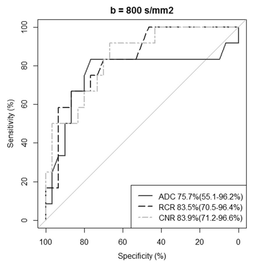
The areas under the ROC curves for the CNR (86.4% [95% confidence interval (CI) 77.2–95.6] and 83.9% [95% CI 71.2–96.6] for b = 1000 and 800 s/mm2, respectively) and the RCR (85.3% [95% CI 75.5–94.8] and 83.5% [95% CI 70.5–96.4]) tended to be superior to the ADC (71.1% [95% CI 56.9–85.2] and 75.7% [95% CI 55.1–96.2]; p < 0.05 for CNR vs ADC for b = 1000 s/mm2, but not significant for other parameters) for discrimination of poorly differentiated HCCs.
The ICCs for the RCR (0.93 [95% CI 0.88–0.96] and 0.87 [95% CI 0.77–0.93]) tended to be greater than for the CNR (0.81 [95% CI 0.70–0.88] and 0.63 [95% CI 0.41–0.78]) and the ADC (0.65 [95% CI 0.28–0.82] and 0.75 [95% CI 0.36–0.89]).
Discussion
HCC is known to increase T2 elongation and to restrict diffusion with increasing histological grade.6, 15, 16 The signal intensities of DWI, which are presented as the RCR and the CNR, are influenced by T2 elongation as well as restriction of diffusion. Therefore, the RCR and CNR might be more appropriate for predicting the histological grade of HCC than measurement of the ADC alone.
ROC analyses revealed that the RCR and CNR on b = 1000 s/mm2 tended to be superior to the RCR and CNR on b = 800 s/mm2 for discrimination of pHCCs. Restriction of diffusion is more strongly related to the contrast in DWI on b = 1000 s/mm2 than on b = 800 s/mm2 and is considered to be a more important reflection of histological grade. A previous study that used methods similar to those in the present study showed that the AUCs of ADC on b = 0 and 1500 s/mm2 were greater than those on b = 800–1000 s/mm2 in the present study.6
There are several limitations to this study, in particular its retrospective design and small patient population. Further prospective studies in large cohorts are needed. Another limitation is that the reproducibility of RCR and CNR among the different magnetic field strengths and respiratory motion compensation techniques available is unknown. Further assessment of the reproducibility of RCR and CNR using other measurement methods under various conditions of MRI acquisition might be needed.
Conclusion
This multi-vendor study demonstrated that RCR, CNR, and ADC with a b-value of 800 and 1000 s/mm2 could discriminate HCC grade, and that non-ADC parameters such as RCR and CNR on DWI might be more useful than ADC for predicting pHCC.Acknowledgements
No acknowledgement found.References
1. Schwarz RE, Smith DD. Trends in local therapy for hepatocellular carcinoma and survival outcomes in the US population. Am J Surg. 2008; 195: 829-836.
2. Zhou L, Rui JA, Wang SB, Chen SG, Qu Q, Chi TY, et al. Outcomes and prognostic factors of cirrhotic patients with hepatocellular carcinoma after radical major hepatectomy. World J Surg. 2007; 31: 1782-1787.
3. Zhou L, Rui JA, Wang SB, Chen SG, Qu Q, Chi TY, et al. Factors predictive for long-term survival of male patients with hepatocellular carcinoma after curative resection. J Surg Oncol. 2007; 95: 298-303.
4. Haratake J, Takeda S, Kasai T, Nakano S, Tokui N. Predictable factors for estimating prognosis of patients after resection of hepatocellular carcinoma. Cancer. 1993; 72: 1178-1183.
5. Chang WC, Chen RC, Chou CT, Lin CY, Yu CY, Liu CH, et al. Histological grade of hepatocellular carcinoma correlates with arterial enhancement on gadoxetic acid-enhanced and diffusion-weighted MR images. Abdom Imaging. 2014; 39: 1202-1212. 6.
Iwasa Y, Kitazume Y, Tateishi U, Saida Y, Ban D, Tanabe M, et al., Hepatocellular Carcinoma Histological Grade Prediction: A Quantitative Comparison of Diffusion-Weighted, T2-Weighted, and Hepatobiliary-Phase Magnetic Resonance Imaging. J Comput Assist Tomogr. 2016; 40: 463-470.
7. Le Moigne F, Boussel L, Haquin A, Bancel B, Ducerf C, Berthezène Y, et al. Grading of small hepatocellular carcinomas (≤2 cm): correlation between histology, T2 and diffusion-weighted imaging. Br J Radiol. 2014; 87: 20130763.
8. An C, Park MS, Jeon HM, Kim YE, Chung WS, Chung YE et al. Prediction of the histopathological grade of hepatocellular carcinoma using qualitative diffusion-weighted, dynamic, and hepatobiliary phase MRI. Eur Radiol. 2012; 22: 1701-1708.
9. Saito K, Moriyasu F, Sugimoto K, Nishio R, Saguchi T, Akata S, et al. Histological grade of differentiation of hepatocellular carcinoma: comparison of the efficacy of diffusion-weighted MRI with T2-weighted imaging and angiography-assisted CT. J Med Imaging Radiat Oncol. 2012; 56: 261-269.
10. Nakanishi M, Chuma M, Hige S, Omatsu T, Yokoo H, Nakanishi K, et al. Relationship between diffusion-weighted magnetic resonance imaging and histological tumor grading of hepatocellular carcinoma. Ann Surg Oncol. 2012; 19: 1302-1309.
11. Nishie A, Tajima T, Asayama Y, Ishigami K, Kakihara D, Nakayama T, et al. Diagnostic performance of apparent diffusion coefficient for predicting histological grade of hepatocellular carcinoma. Eur J Radiol. 2011; 80: e29-33.
12. Heo SH, Jeong YY, Shin SS, Kim JW, Lim HS, Lee JH et al. Apparent diffusion coefficient value of diffusion-weighted imaging for hepatocellular carcinoma: correlation with the histologic differentiation and the expression of vascular endothelial growth factor. Korean J Radiol. 2010; 11: 295-303.
13. Nasu K, Kuroki Y, Tsukamoto T, Nakajima H, Mori K, Minami M. Diffusion-weighted imaging of surgically resected hepatocellular carcinoma: imaging characteristics and relationship among signal intensity, apparent diffusion coefficient, and histopathologic grade. Am J Roentgenol. 2009; 193: 438-44.
14. Muhi A, Ichikawa T, Motosugi U, Sano K, Matsuda M, Kitamura T, et al., High-b-value diffusion-weighted MR imaging of hepatocellular lesions: estimation of grade of malignancy of hepatocellular carcinoma. J Magn Reson Imaging. 2009; 30: 1005-1011.
15. Ebara M, Fukuda H, Kojima Y, Morimoto N, Yoshikawa M, Sugiura N, et al. Small hepatocellular carcinoma: relationship of signal intensity to histopathologic findings and metal content of the tumor and surrounding hepatic parenchyma. Radiology. 1999; 210, 81-88.
16. Kadoya M, Matsui O, Takashima T, Nonomura A. Hepatocellular carcinoma: correlation of MR imaging and histopathologic findings. Radiology. 1992; 183: 819-825.
Figures