1664
DWI assessment of the optic nerve and chiasma of acute optic neuritis: Advantages in field-of-view optimized and constrained undistorted single shot (FOCUS) method1radiology department, General Hospital of the People’s Liberation Army, Beijing, China
Synopsis
In the current study, we evaluated the performance of the field-of-view optimized and constrained undistorted single shot (FOCUS) DWI in assessing the optic nerve and chiasma abnormalities of acute optic neuritis. Visual assessment was obtained for the FOCUS-DWI and the conventional-DWI (c-DWI). We found that FOCUS-DWI provided better visual assessments of the optic nerve and chiasma abnormalities in acute optic neuritis (AON), with much reduced blurring effects and geometric deformations. It might indicated that the FOCUS-DWI would improve the diagnostic accuracy and prognosis evaluation in AON.
Purpose
To evaluate the performance of field-of-view (FOV) optimized and constrained undistorted single shot (FOCUS) diffusion-weighted imaging (DWI; FOCUS-DWI) in assessing the optic nerve and chiasma abnormalities of acute optic neuritis (AON), and make comparison with the performance of conventional DWI(c-DWI).Introduction
Methods
This study was approved by the local institutional review board. 36 patients clinically diagnosed as having new-onset acute optic neuritis (AON; within 1 month from onset) were included in the current study. MRI scanning was performed on a 3-T MR scanner (Discovery750, GE Healthcare, Waukesha, WI, USA) with a 32-channel head coil. The c-DWI was performed with field-of-view (FOV) of 18.0 ×18.0 cm, image matrix was 160 × 160 and the resolution of 1.1 × 1.1 × 3.0 mm. The field-of-view in the FOCUS-DWI was reduced to 19.0 × 8.0 cm with matrix of 160 × 80, resulting in the same resolution as the c-DWI. Two radiologists with more than 5 years of experience in reading MRI of the orbits, blindly and independently evaluated all images on a standard ADW4.6 workstation (GE Healthcare). Visual assessment including overall image quality, lesion conspicuity, artifacts, distortion and degree of image blurring were rated on a five-point Likert scale on the c-DWI and the FOCUS-DWI images of the AON patients. All the results from visual assessments were compared using the Wilcoxon-Mann-Whitney test. Findings were reported as means ± standard deviations. The level of inter-observer agreement for each assessed qualitative parameter was evaluated by Cohen’s Kappa (k).Results
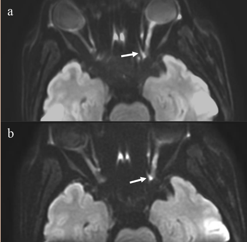
The overall image quality (P < 0.0001) and lesion conspicuity (P < 0.0001) of the AON patients was found to be significantly better on the FOCUS-DWI than the c-DWI. It was also observed that the artifacts (P < 0.0001), distortions (P < 0.0001), and image blurring (P < 0.0001) were all less severe on FOCUS-DWI than the c-DWI. The results of the qualitative evaluations are presented in Table 1. We also illustrated that the artifacts in the FOCUS-DWI images and the c-DWI images for a 42-year-old female patient with optic neuritis in Fig 1. Inter-observer agreement between the two readers was substantial for all the image evaluation categories.Discussion
The results of the present study revealed that when compared visually at the same resolution, FOCUS-DWI was superior to c-DWI imaging in all the visual assessments studied. Demyelinative optic neuritis is the most common cause of non-glaucomatous optic neuropathy in younger adults7. There have been a number of studies reporting that DWI could provide a promising tool to improve the diagnostic performance of AON1, 3, 8. However, the most wildly used approach of c-DWI was likely to produce magnetic susceptibility artifacts, chemical shift artifacts, and low image quality1, 9. On the other hand, the FOCUS-DWI applies a 2-dimensional spatially selective echo-planar radiofrequency excitation pulse and a 180° refocusing pulse to reduce the FOV in the phase-encode direction, achieving high spatial resolution images with less distortion.Conclusions
In the current study, it was found that FOCUS-DWI provided better visual assessments of the optic nerve and chiasma abnormalities in AON, with much reduced blurring effects and geometric deformations.Acknowledgements
No acknowledgement found.References
1. Wan H, Sha Y, Zhang F, Hong R, Tian G, Fan H (2016) Diffusion-weighted imaging using readout-segmented echo-planar imaging, parallel imaging, and two-dimensional navigator-based reacquisition in detecting acute optic neuritis. J Magn Reson Imaging 43:655-660
2. McKinney AM, Lohman BD, Sarikaya B, Benson M, Lee MS, Benson MT (2013) Accuracy of routine fat-suppressed FLAIR and diffusion-weighted images in detecting clinically evident acute optic neuritis. Acta Radiol 54:455-461
3. Jeong HK, Dewey BE, Hirtle JA et al (2015) Improved diffusion tensor imaging of the optic nerve using multishot two-dimensional navigated acquisitions. Magn Reson Med 74:953-963
4. Dong H, Li Y, Yu K, Li H (2016) Comparison of image quality and application values on different field-of-view diffusion-weighted imaging of breast cancer. Acta Radiol 57:19-24
5. Feng Z, Min X, Sah VK et al (2015) Comparison of field-of-view (FOV) optimized and constrained undistorted single shot (FOCUS) with conventional DWI for the evaluation of prostate cancer. Clin Imaging 39:851-855
6. Wheeler-Kingshott CA, Parker GJ, Symms MR et al (2002) ADC mapping of the human optic nerve: increased resolution, coverage, and reliability with CSF-suppressed ZOOM-EPI. Magn Reson Med 47:24-31
7. Onodera M, Yama N, Hashimoto M et al (2015) The signal intensity ratio of the optic nerve to ipsilateral frontal white matter is of value in the diagnosis of acute optic neuritis. Eur Radiol. 10.1007/s00330-015-4114-4
8. Dowell NG, Jenkins TM, Ciccarelli O, Miller DH, Wheeler-Kingshott CA (2009) Contiguous-slice zonally oblique multislice (CO-ZOOM) diffusion tensor imaging: examples of in vivo spinal cord and optic nerve applications. J Magn Reson Imaging 29:454-460
9. Bender B, Heine C, Danz S et al (2014) Diffusion restriction of the optic nerve in patients with acute visual deficit. J Magn Reson Imaging 40:334-340
Figures
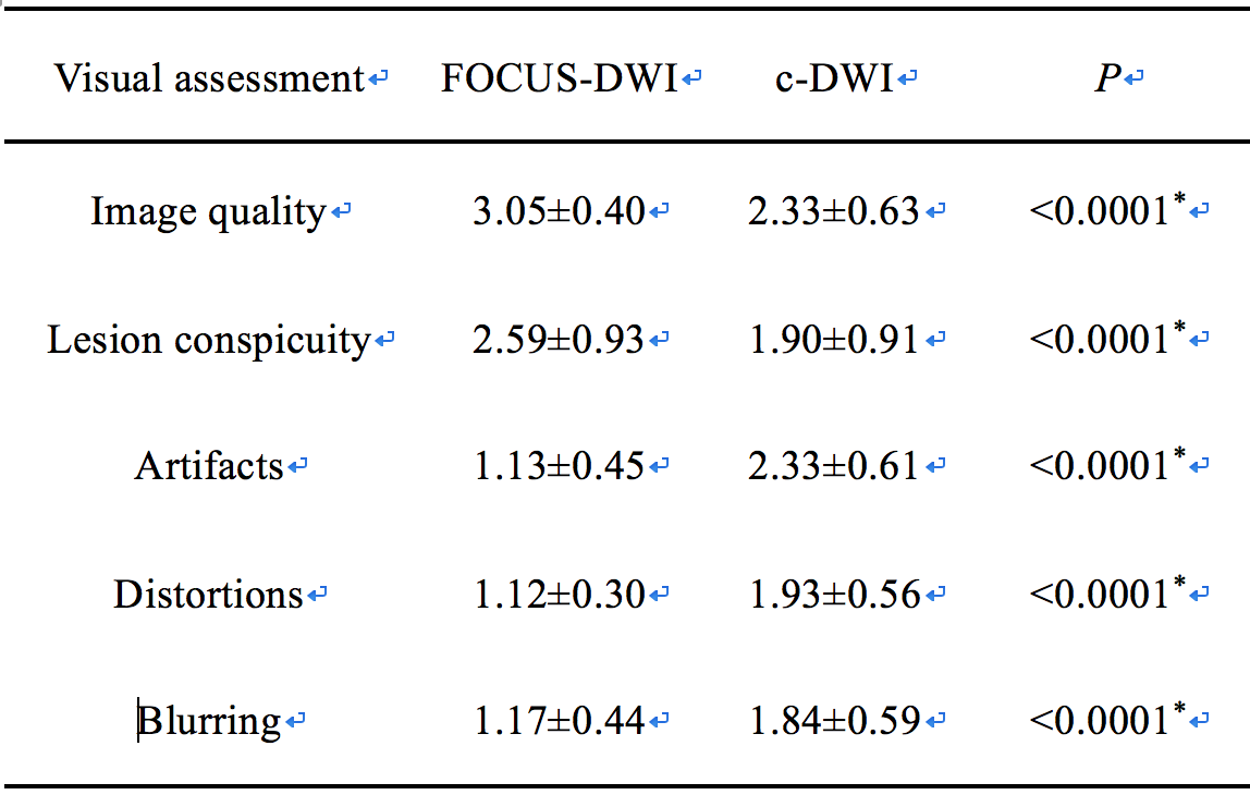
Table.1 Qualitative evaluation of FOCUS-DWI and c-DWI on five-point Likert scales
Data are mean ± standard deviations (range in brackets)
Five-point Likert scale from 0 to 4
* indicated P<0.001 in the Wilcoxon-Mann-Whitney test