1644
Removal or correction of volumes affected by bulk motion: impact on DTI and NODDI metrics1CSIRO, Brisbane, Australia, 2University of Newcastle, Newcastle, Australia, 3The University of Queensland, Brisbane, Australia
Synopsis
In difficult patient populations, the interleaved acquisition of diffusion weighted volumes often leads to images that are not self-consistent due to movement. Here, we investigated the effect of removing or correcting volumes with movement artefacts on the DTI measures FA and MD, as well as on NODDI measures. While removal of affected volumes is typically used, we found that a simple correction strategy leads to markedly lower bias and variability in all diffusion measures. Data that may need to be rejected entirely if volume removal is used, may be salvaged if correction is used.
Introduction
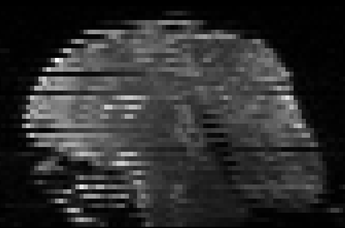
Bulk head motion is frequently observed in diffusion MRI of difficult patient populations, such as neonates. Bulk head motion during the interleaved acquisition of diffusion-weighted volumes can result in volumes that are not self-consistent (Figure 1). Typically, affected volumes are removed before subsequent processing. The impact of removing volumes (and their associated diffusion directions) has been studied previously for DTI data1. Here, we investigate the impact on NODDI measures, and determine whether correcting rather than removing affected volumes improves the reliability of calculated FA and MD, and NODDI measures.Methods
Diffusion MRI data (30 directions at b=1000s/mm2 [DTI] and 64 directions at b=2000s/mm2 [HARDI]) of preterm infants scanned at term-equivalent age were used in this study. One participant without visible bulk head motion in any volumes was selected for simulation studies. Preprocessing included correction for head movement between volumes with adjustment of the b-matrix and correction of susceptibility distortions using a field map.
The impact of removing or correcting volumes was simulated by removing/correcting between 1 and 23/59 (DTI/HARDI) randomly selected volumes (i.e. retaining at least 6 volumes; 100 random realisations each). Correction of volumes was performed by splitting volumes into two subvolumes consisting of only odd and even slices, respectively, using cubic spline interpolation to preserve slice thickness. These volumes were used to replace the affected volume.
Diffusion data were upsampled by a factor of 2. The diffusion tensor and, subsequently, maps of FA and MD were calculated using MRtrix32, separately for DTI and HARDI data. NODDI maps of intra-cellular volume fraction (ICVF), isotropic volume fraction (IsoVF), and orientation dispersion (OD) were calculated using AMICO3 using the combined DTI and HARDI data.
The FA maps of DTI and HARDI acquisitions were averaged, and non-linearly registered to the JHU neonatal template4. The JHU neonatal atlas was then transformed into subject space. The left posterior limb of the internal capsule (PLIC) was used as an example region of interest. Average values of FA, MD, ICVF, IsoVF, and OD were calculated for this region.
Results
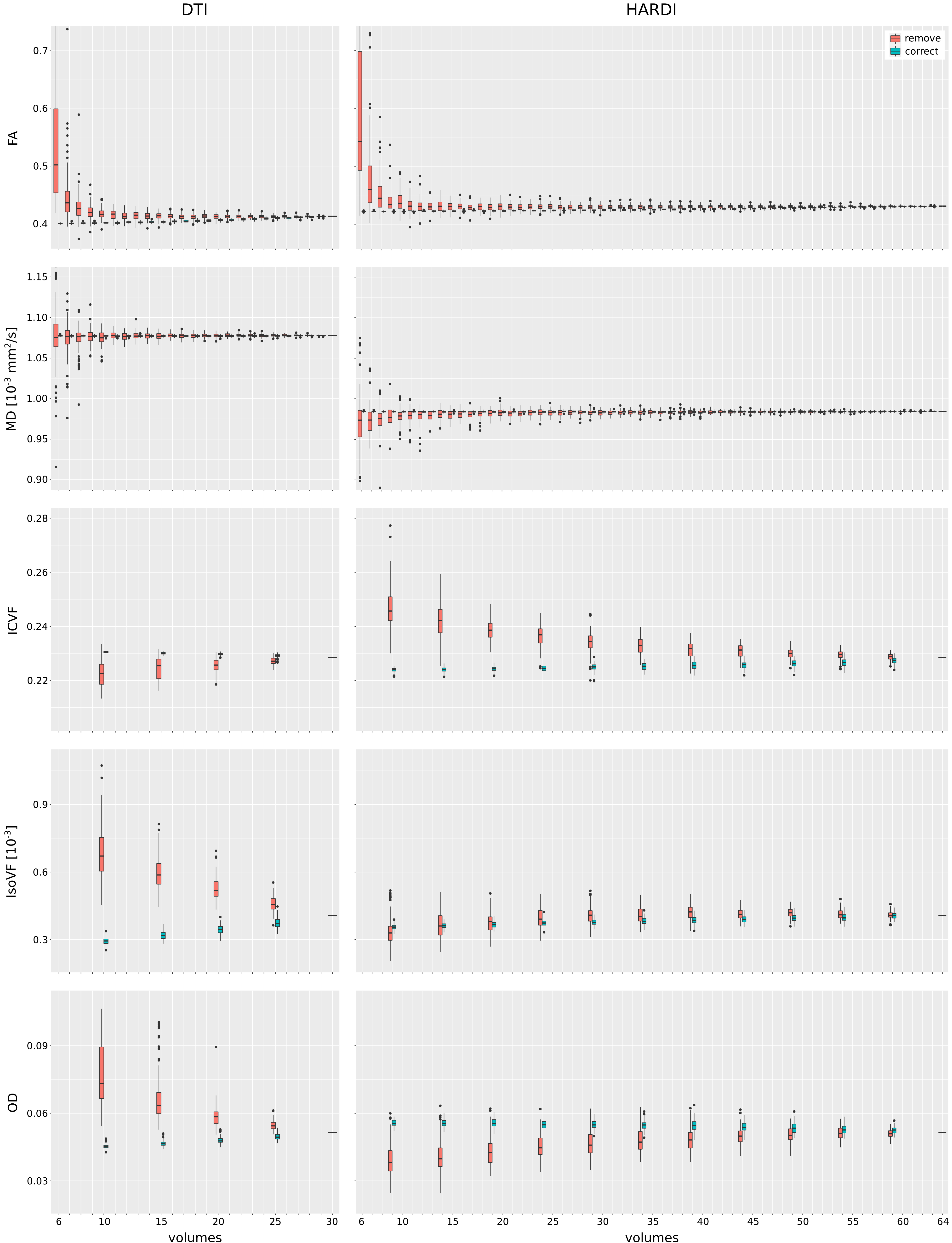
Impact on DTI measures (Figure 2, top): Removal of an increasing number of volumes resulted in a positive bias and increasing variability in FA. Patterns of FA changes were similar for DTI and HARDI data. Variability of MD increased with an increasing number of removed volumes; for HARDI data, but not for DTI data, a negative bias was observed. In contrast, correction of volumes led to a small negative bias in FA, and small positive bias in MD. Variability of FA and MD was markedly reduced for correction compared to removal.
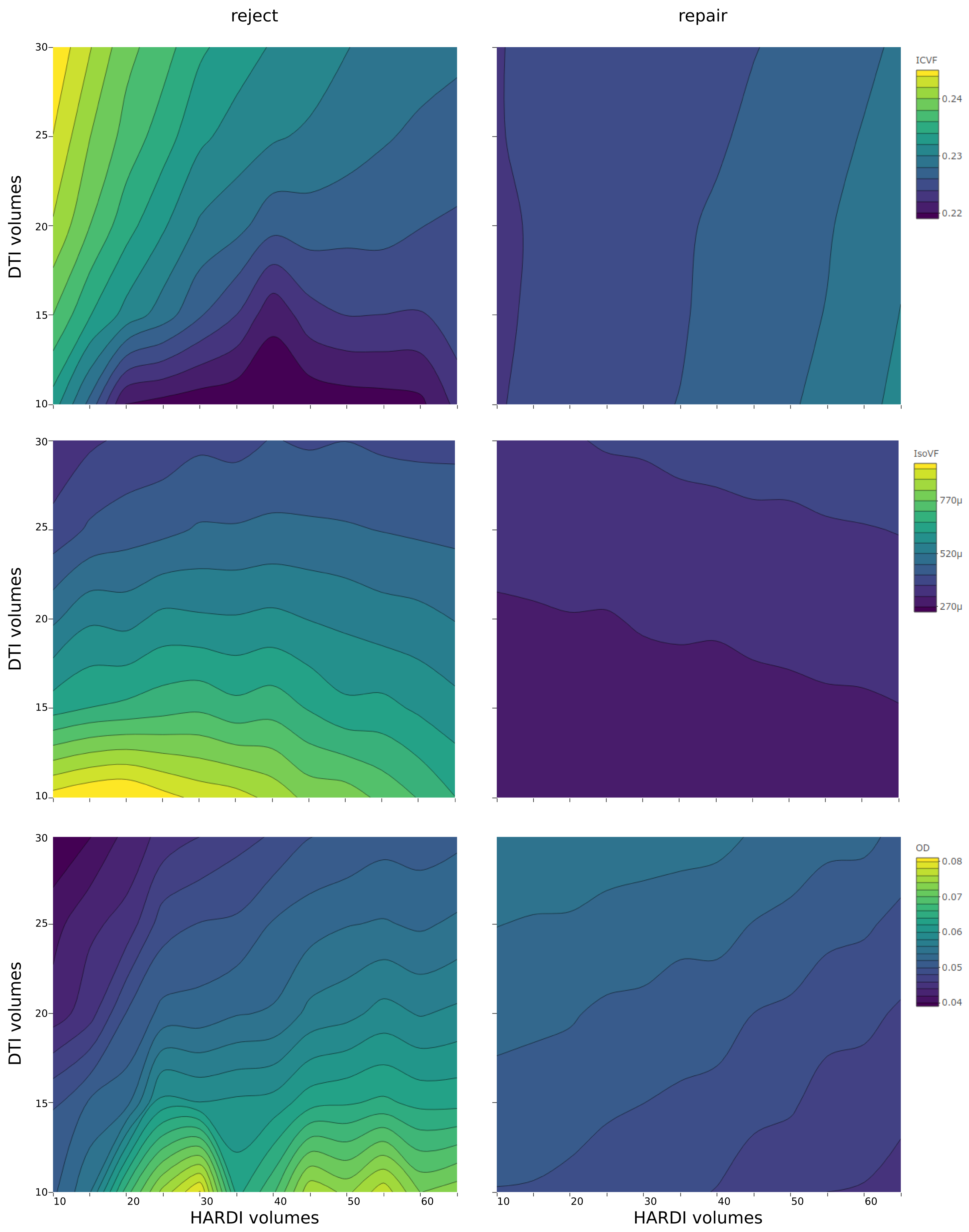
Impact on NODDI measures (Figure 2, bottom; Figure 3): As with DTI measures, correction of volumes had a lower impact on NODDI measures of ICVF, IsoVF, and OD than their removal: both the observed bias, and the observed variability were markedly reduced when correction was used. Removal of low b-value (DTI) volumes led to a negative bias in ICVF, and a positive bias in IsoVF and OD. In contrast, removal of high b-value (HARDI) volumes led to a positive bias in ICVF and a negative bias in IsoVF and OD. Correction of low b-value (DTI) volumes led to a smaller positive bias in ICVF, and smaller negative bias in IsoVF and OD. Correction of high b-value (HARDI) volumes led to a smaller negative bias in ICVF and IsoVF, and smaller positive bias in OD.
Discussion
This study demonstrates that even a simple correction strategy for volumes affected by motion can result in marked improvements in the reliability of obtained diffusion measures. When affected volumes are removed, the large bias and variability of resulting measures often means that the entire sequence may need to be rejected; however, the data may be salvaged if correction is used. Future work will determine whether bias and variability can be further reduced by incorporating information from all diffusion volumes rather than only the affected diffusion volume.Acknowledgements
This work was supported by grants from the Cerebral Palsy Alliance Research Foundation, the Financial Markets Foundation for Children, the Queensland Government (Smart State; Health Practitioner Stimulus Grant), the Australian National Health and Medical Research Council and the Royal Brisbane and Women’s Hospital. The authors received funding from The University of Queensland, the Queensland Government, and the National Health and Medical Research council.References
1. Chen, Y., Tymofiyeva, O., Hess, C. P., & Xu, D. (2015). Effects of rejecting diffusion directions on tensor-derived parameters. NeuroImage, 109, 160-70.
2. Tournier, J., Calamante, F., & Connelly, A. (2012). MRtrix: Diffusion tractography in crossing fiber regions. International Journal of Imaging Systems and Technology, 22(1), 53-66
3. Daducci, A., Canales-Rodríguez, E. J., Zhang, H., Dyrby, T. B., Alexander, D. C., & Thiran, J. P. (2015). Accelerated microstructure imaging via convex optimization (AMICO) from diffusion MRI data. NeuroImage, 105, 32-44
4. Oishi, K., Mori, S., Donohue, P. K., Ernst, T., Anderson, L., Buchthal, S., . . . Chang, L. (2011). Multi-contrast human neonatal brain atlas: Application to normal neonate development analysis. NeuroImage, 56(1), 8-20
Figures