1642
2D Single-Shot Radial Diffusion-Weighted Imaging free of geometric distortion and optimization of SNR using Variable Flip-Angle and Random View-Ordering1Utah Center for Advanced Imaging and Research, University of Utah, Salt Lake City, UT, United States, 2Department of Bioengineering, University of Utah, Salt Lake City, UT, United States, 3Radiology and Imaging Sciences, University of Utah, Salt Lake City, UT, United States
Synopsis
The 2D ss-DWEPI is routinely used for in-vivo DW imaging, because of its immunity to motion-induced artifact, but prone to susceptibility-induced geometric distortion. We present a novel DWI technique using single-shot radial imaging, which produces DW images with minimal geometric distortion, no motion artifact, and with optimized SNR and reduced effect of undersampling radial streak artifact. Variable-flip angle (VFA) and random-view ordering (RVO) were implemented to improve the SNR and reduce the geometric distortion, respectively.
Introduction
2D single-shot diffusion-weighted EPI (2D ss-DWEPI) is commonly used for diffusion MRI, because of its immunity to motion-induced artifact. However, DWI using the 2D ss-DWEPI is subject to severe geometric distortion at/near the large field gradient, such as air-tissue and bone-tissue interfaces [1]. Therefore, the application of 2D ss-DWEPI is limited to intracranial brain, far from the sinus and temporal bones. 2D single-shot radial DWI (2D ss-rDWI), too, is insensitive to motion artifact due to heavy k-space sampling at the k-space center [2] and with acquisition of complete k-space coverage after a single diffusion-preparation [3], but with low SNR. In this abstract, we present 2D ss-rDWI that produces DW images with optimized signal-to-noise ratio (SNR) and reduced reconstruction-related distortion using variable flip-angle (VFA) and random view-ordering (RVO), respectively. We also compare the performance of 2D rDWI using VFA and RVO to constant flip-angle (CFA) and smooth view-ordering (SVO).Methods
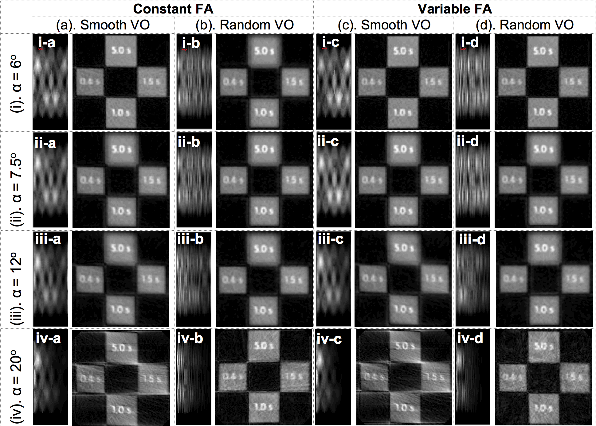
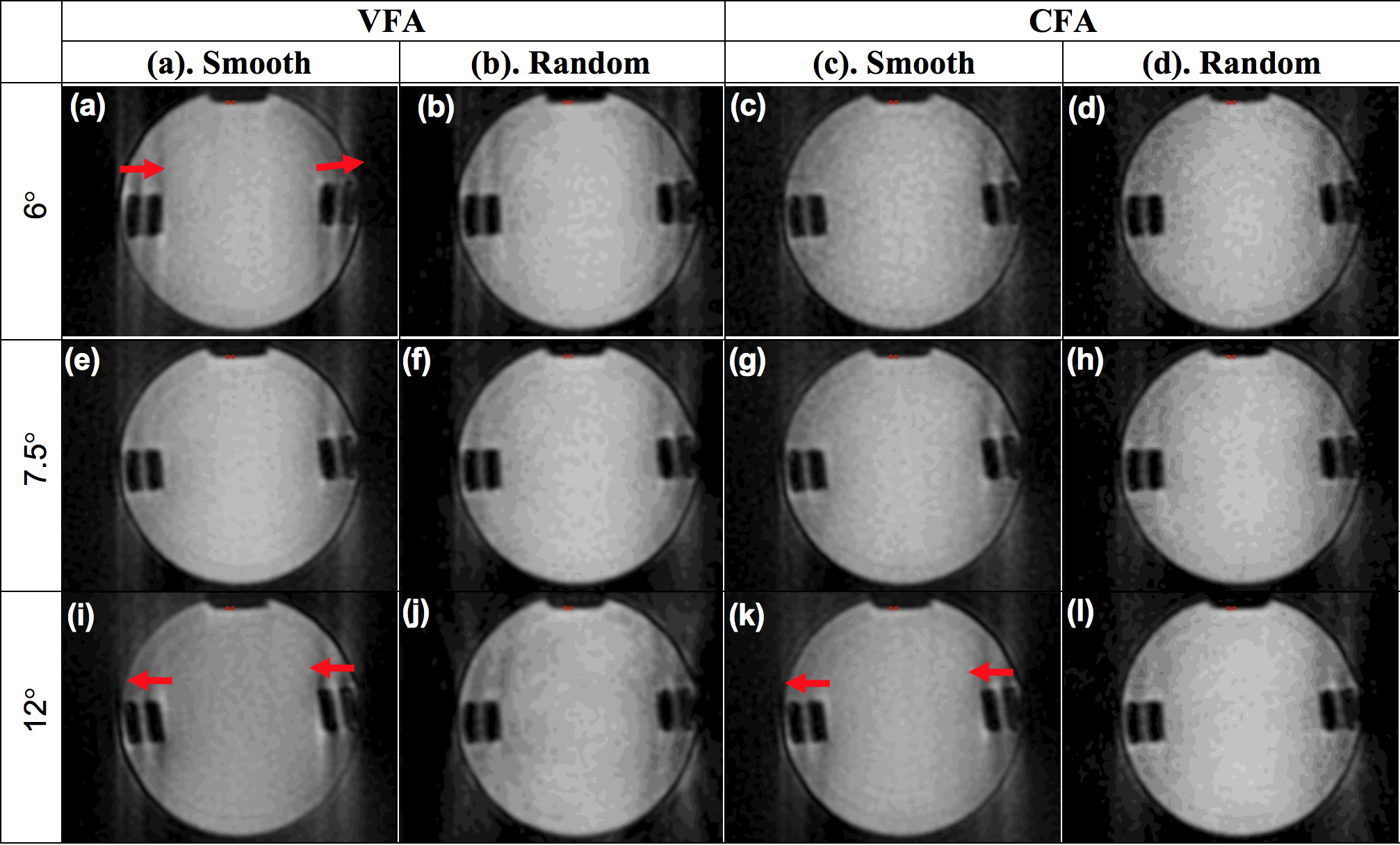
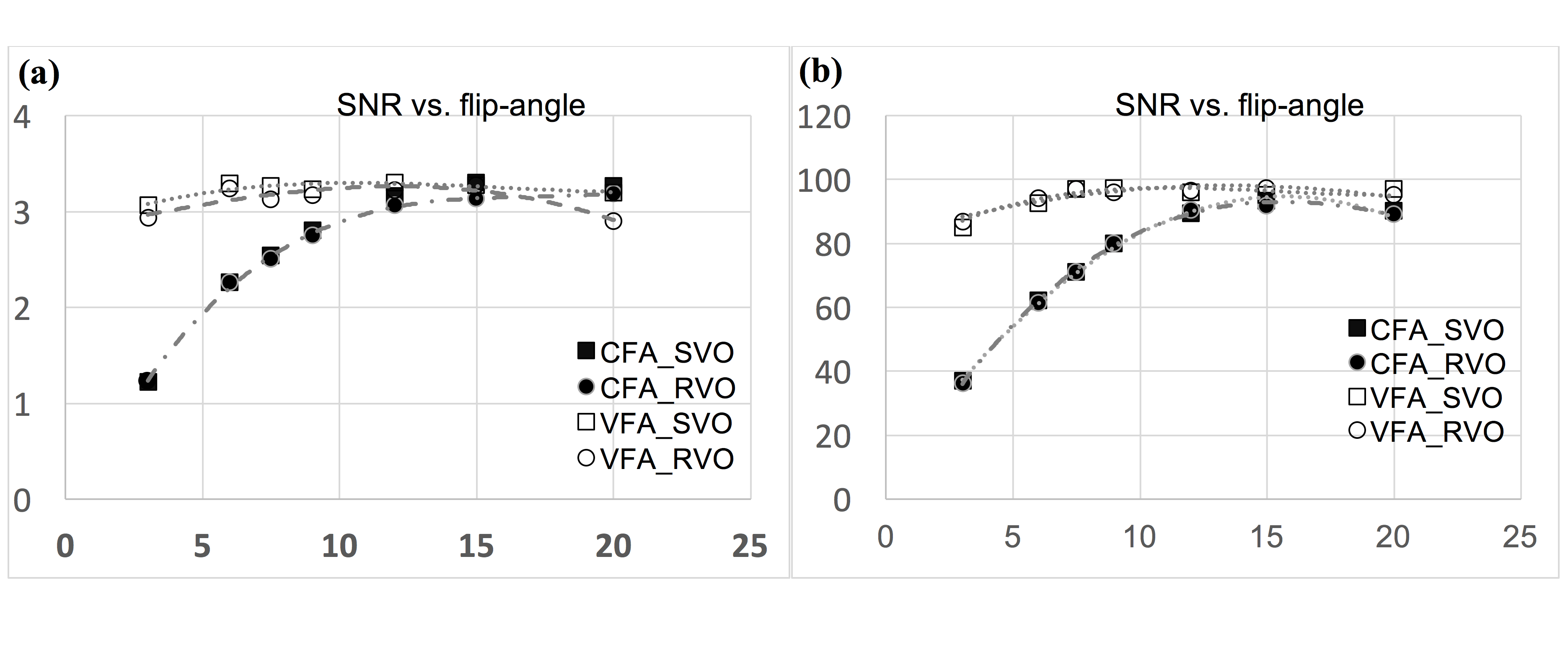
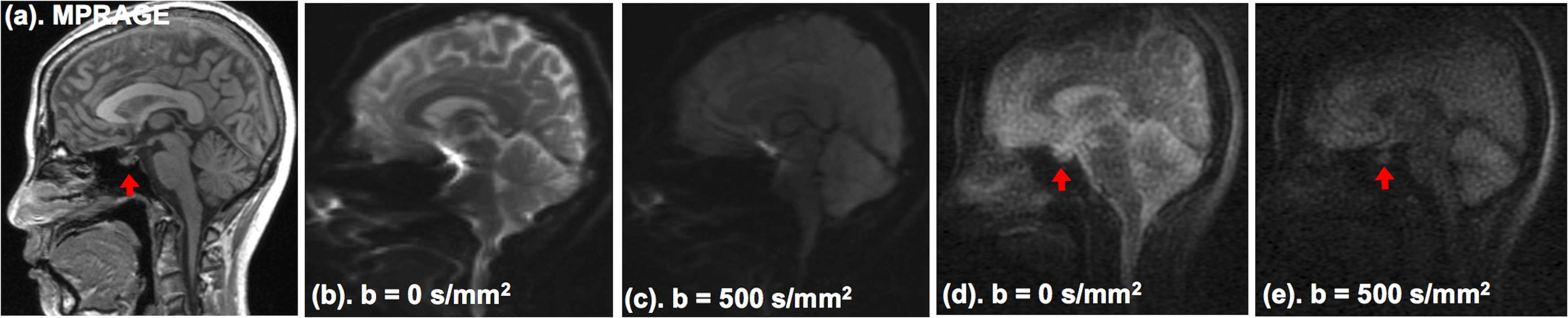
Diffusion-preparation module is developed and applied prior to the data acquisition. Because the diffusion-prepared longitudinal magnetization is measured over a multiple RF excitations for 48 radial spokes in each shot, images are subject to low signal-to-noise ratio (SNR). We used VFA and RVO to optimize the SNR and to reduce the sampling-related artifact, respectively. Flip-angle αn of the nth RF pulse for VFA is calculated using αn = arctan(sin αn+1·e-τ/T1) for a given T1, with αlast = 90o [4]. For each technique, numerical simulation was performed using a homemade software, programmed using Python language. MRI experiments were performed at 3T MRI system (Prisma, Siemens Medical Solutions, Erlangen, Germany). Phantom MRI experiment was performed to mimic the numerical simulation for comparison. 2D ss-rDWI was then applied for DWI of human brain with eight shots of 48 radial views, TR = 4.0 s, TE = 37 ms, 2x2x4 mm3 resolution, and b = 0 and 500 sec/mm2, and acquisition time of 32 sec for each b-value. DW images were also obtained using 2D ss-DWEPI with similar imaging parameters.Results
The simulations and the MR experimental data presented matching results. Both simulated and MR phantom data revealed insensitivity to magnetic susceptibility-induced geometric distortion when radial view order is randomized in Fig. 1(i-iv)b, Fig. 1(i-iv)d, and Fig. 2(b,d,f,h,j,l). In contrast, SVO proved to be highly sensitive to the geometric distortion as shown in Fig. 1(i-iv)a, Fig. 1(i-iv)c, and Fig. 2(a,c,e,g,i,k). Furthermore, the simulated and experimental MR data using VFA scheme exhibit higher SNR than its CFA counterpart, by consuming the entire diffusion-prepared magnetization in each single-shot, as shown in Fig. 3. Fig. 4(b,c) illustrate DW images using 2D ss-DWEPI with severe geometric distortion for tissues surrounding the sinus space, including the pituitary gland. This geometric distortion was absent in our 2D ss-rDWI images in Fig. 4(d,e) where the pituitary gland retained its visual structure, although with low SNR.Conclusion
We presented a 2D ss-rDWI technique with radial k-space acquisition that produces DWI without motion-induced artifact and geometric distortion, and with reduced streak artifacts using variable flip angle and random view ordering. Preliminary application of this technique on skull base of a volunteer demonstrated a great potential for DWI of other organs, including, but not limited to, upper liver, prostate, and skull-base brain.Acknowledgements
This work is supported by a Research Grant from National Multiple Sclerosis Society (NMSS).References
[1]. Sapkota N, et.al., MRM 2016; 77(6): 2167-2173.
[2]. Bernstein M, et.al. Handbook of MRI Pulse Seuqneces 2005.
[3]. Saritas E, et.al., ISMRM 2008; p. 760.
[4]. Jeong EK, et.al., MRM 2006; 56:1173–1181.
Figures