1638
Distortion Correction using Reverse Polarity Gradient Method: Algorithm Optimization for Prostate Imaging using a Hybrid Weighting MetricMaggie M Fung1, Pauline Worters2, Ek Tsoon Tan3, Arnaud Guidon4, and Ersin Bayram5
1Applications & Workflow, GE Healthcare, New York, NY, United States, 2Applications & Workflow, GE Healthcare, Menlo Park, CA, United States, 3Global Research Center, GE, Niskayuna, NY, United States, 4Applications & Workflow, GE Healthcare, Boston, MA, United States, 5Applications & Workflow, GE Healthcare, Houston, TX, United States
Synopsis
Prostate Diffusion Weighted Echo Planar imaging (DW-EPI) routinely suffers from nonlinear geometric distortion due to B0 inhomogeneity. Although reverse phase-encoding polarity-based distortion correction method works well in the brain, the same technique causes artifacts in prostate DWI due to the low SNR nature of body DWI scans, and the inconsistency of image content between the reverse and forward polarity images. In this study, we showed that a hybrid weighting metric method could improve the distortion correction performance in prostate DWI.
Purpose
Diffusion Weighted Echo Planar imaging (DW-EPI) routinely suffers from nonlinear geometric distortion due to B0 inhomogeneity1. Artifacts are most pronounced at air-tissue interfaces, such as the rectum, in prostate diffusion images. One of the distortion correction methods, reverse phase-encoding polarity gradient (RPG) method2, has been successfully applied to brain DWI. However, directly applying the same technique to prostate DWI resulted in wavy artifacts(Fig.1). In this study, we proposed a hybrid weighting metric as part of the optimization to improve the RPG performance.Theory
Standard RPG method uses a multi-tier biconjugate gradients stabilized method (Bi-CGSTAB)3 to perform non-linear registration on the reverse and forward images to find the undistorted solution (Fig 2). However, while this method worked well in brain DWI with high SNR, this method resulted in wavy artifacts and over-correction when applied to body imaging. The wavy artifact was caused by the incorrect attempt of the algorithm to match the noise speckles, and the over-correction was caused by the anatomical mis-match between the reverse and forward images (Fig 1). We had explored using a weak diffusion gradient to remove this effect, however, even at B=50, there were still some differences in the vessel appearances, so that alone could not fully address the artifacts. As a result, we introduced a metric to represent the pixel-by-pixel similarity between the reverse and forward image sets and used this metric as a weighting factor during the Bi-CGSTAB optimization. The rationale was that if the pixels do not correlate well (due to anatomical differences or due to noise), the algorithm should weight less in that area during the cost function minimization. However, in regions where the B0 inhomogeneity was severe and not surrounded by other high SNR anatomies (e.g. rectal wall), since the reverse and forward images will not match due to distortion, those areas were not heavily weighted using the fore-mentioned metric. As a result, we had to rely on a second metric (defined by the shim volume location) to ensure those areas were included in the registration. The optimized algorithm flow chart is shown in figure 2.Method
Axial DWI were performed on 8 healthy volunteers on a 3T 70cm bore scanner (Signa Premier, GE Healthcare, USA) using the GEM anterior andposterior array. DWI parameters were: FOV:24cmx24cm, Matrix: 120x120, TR/TE:3866ms/53.3ms, single spin echo, BW:250kHz, slice thickness:4mm, # slices:22, Fat Sat, b-value=0s/mm2(1 nex), 50s/mm2 (4 nex), 800s/mm2 (12nex), diffusion encoding: ALL, anterior and posterior sat bands, scan time: 3:17min. In 5 cases, reference T2 FSE was also acquired with these parameters: FOV:24cmx24cm, Matrix: 256x224, TR/TE:4985ms/77.6ms, BW:31.3kHz, Slice thickness:4mm, # slices:22, nex: 3, scan time: 3:05min. We compared the undistorted image qualitatively in all 8 cases. In 5 cases where T2 FSE reference were acquired, mutual information(MI) between the T2 and DWI image (standard vs new RPG) were calculated globally (in the entire slice) and locally (in the prostate area) and compared using a two-sample t-test.Results and Discussions
Qualitative visual comparison between the algorithms showed that wavy artifacts were not observed in the optimized RPG cases. In 7/8 cases, the two algorithms showed similar spatial distortion correction in the prostate and seminal vesicle areas, while in 6/8 cases, the two algorithms showed similar distortion correction rectal wall area. Fig.3 shows a comparison between no RPG, original and optimized RPG algorithm. The optimized RPG algorithm corrected for the prostate and rectum but did not over-correct the vessels. The wavy artifacts in the original RPG algorithm was especially pronounced at high b-value images, and the optimized RPG algorithm greatly improved that(Fig.4). In the quantitative analysis, the MI metric improved as expected when either of the RPG algorithms were employed (Fig.5). There were no statistical significant differences between the MI metric of the original vs optimized methods (0.4513±0.1055 vs 0.4383±0.1070 (p=0.4286) for global MI, 0.8225±0.1588 vs 0.8151±0.1641 (p=0.7638) for local MI). In general, the new algorithm maintained the correction performance at the prostate and does not generate wavy artifacts.Conclusion
We have demonstrated an optimized RPG technique in prostate imaging using a hybrid weighting metric. This approach considered the SNR and similarity between the forward and reverse images and therefore provided a more constrained solution to the distortion correction optimization. Future direction will include expanding this to other anatomies such as breast and liver.Acknowledgements
The author acknowledged Andres Dales (UCSD) for the useful discussion.References
[1] Bernstein, et al. 2004b. Handbook of MRI Pulse Sequences.
[2] Holland, Dale et al, NeuroImage 50 (2010) 175–183
[3] van der Vorst, et al, Cambridge University Press, Cambridge. (April 2003).
Figures
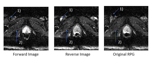
Figure 1. DWI
images with forward & reverse polarity exhibits different vessels
characteristics
(1)
since they are collected at different parts of the cardiac cycle, thus causing
incorrect distortion correction using original RPG algorithm. Also, original
RPG algorithm also attempts to “track the noise speckles” in lower SNR prostate DWI regions (2).
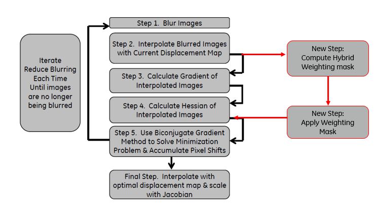
Figure 2. Original
RPG algorithm (in black boxes) and additional steps employed in optimized RPG
algorithm for prostate (in red boxes)
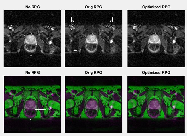
Figure 3. Top
row are DWI images, and bottom row are fused DWI (purple) and T2 (green)
images. Both original RPG & optimized RPG algorithm corrects the distortion
at the prostate & rectal wall (single arrow). While original RPG introduces
wavy artifacts at the vessels (double arrow), optimized RPG does not
overcorrect in those area due to a lower weighting in these area during the Bi-CGSTAB
optimization.
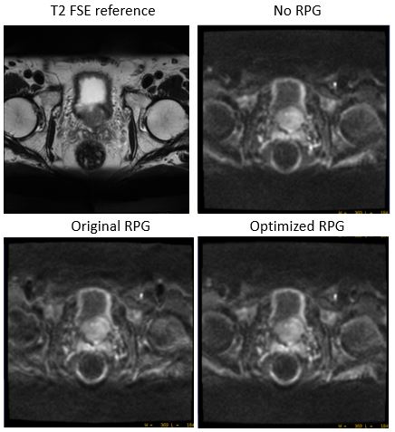
Figure 4. The
correction error in original RPG algorithm propagates to high b value image and
significantly degrade the image quality (wavy artifacts and haziness over the entire image). The optimized RPG
method does not suffers from the same drawback.
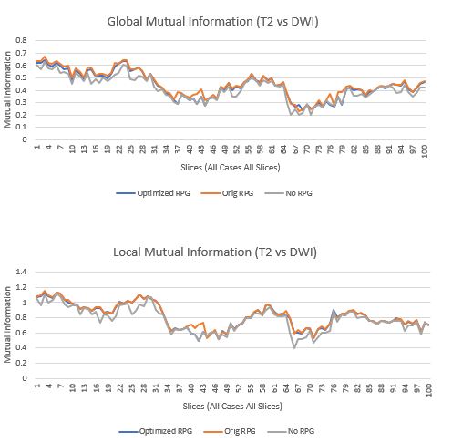
Figure 5. Global mutual information & local
mutual information (only at shim ROI) between T2 FSE reference and DWI image
for all slices in all cases. Optimized and original RPG exhibits similar mutual
information trends, and exhibits higher mutual information compared to no RPG
correction. Local mutual information is in general higher because in some T2
cases, sat bands are not employed, leading to a lower mutual information,
compare to DWI with sat bands employed.