1627
Improvement of diffusion-weighted image quality by iShim toward realization of cervical spinal cord region QSI1Medical Scanning Tokyo, Tokyo, Japan, 2Central Institute for Experimental Animals, Japan, Tokyo, Japan, 3Department of Orthopaedic Surgery, Keio University School of Medicine, Tokyo, Japan, 4MR Research & Collaboration Dpt., Diagnostic Imaging Business Area, Siemens Healthcare K.K., Tokyo, Japan, 5Siemens Healthcare GmbH, Erlangen, Germany, Erlangen, Germany
Synopsis
Herein, we adopted diffusion-weighted imaging (DWI) with a high fat suppression effect and high signal-to-noise ratio (SNR) in the cervical region, where magnetic field inhomogeneity may occur, using integrated slice-by-slice shimming (iShim), which improves static magnetic field (B0) shimming accuracy. We examined spinal cord SNR and standard deviation in healthy volunteers and performed cervical DWI with the conventional B0 shimming method and iShim, respectively. Furthermore, to verify whether short TI inversion recovery (STIR) or water excitation (WE) was appropriate as a fat suppression method, we used DWI with a high SNR at the cervical region by combining iShim with WE.
Introduction
Calculated images based on diffusion-weighted imaging (DWI) obtained by assuming the transition probability density distribution of molecular diffusion, such as fractional anisotropy and apparent diffusion coefficient by normal distribution, maintain a sufficient signal-to-noise ratio (SNR) with high b-values to improve the accuracy of the analysis result1,2. However, even if the average number is increased, which is an imaging parameter affecting SNR, it cannot be expected to greatly improve the accuracy of the calculated image. Therefore, improving the SNR is not easy. Additionally, because DWI emits a chemical shift artifact (CSA), wherein the phase error is accumulated by continuous magnetic field gradient in the phase direction and fat signal as a spatially shifted image is developed, it is essential to combine fat suppression. In the cervical region, it is difficult to obtain a uniform magnetic field and an effective fat suppression because of its complicated shape. However, in the cervical region and mammary gland, whole-body DWI (WBDWI) examination using DWI combined with short TI inversion recovery (STIR) reportedly improved image quality3-5. We developed integrated slice-by-slice shimming (iShim) to improve image quality of WBDWI, particularly in the cervical region (Fig.1)6,7. Therefore, we investigated whether DWI image quality of the cervical region can be improved using iShim to select a fat suppression method with high SNR other than STIR.
Methods
QSI imaged the cervical region using 3T MRI (MAGNETOM Skyra System, Siemens Healthcare, Erlangen, Germany) and 64-ch head/neck coil (Fig. 2).
For SNR of the spinal cord, we compared the SNR from four patterns combining two B0 shimming methods (3D Shim/iShim) and two fat suppression methods (STIR/WE) using DWI trace images. SNR = √2 × spinal cord signal average value/spinal cord standard deviation (SD) average value. For accuracy of the spinal cord analysis, we compared the SD of the spinal cord from four patterns combining two B0 shimming methods (3D shim/iShim) and two fat suppression methods (STIR/WE) using the QSI kurtosis image. For fat suppression effect of the cervical region, we compared the CSA (total signal value of fat suppression remaining in the cervical region) to patterns combining iShim and two B0 shimming methods (3D Shim/iShim) using the DWI trace images. All investigations included 11 healthy volunteers.
Results
The spinal cord SNR using the DWI trace image was higher in the WE group than in the STIR group (P < 0.05) (Fig. 3).
The SD of the spinal cord signal using the QSI kurtosis image was lower in the WE group than in the STIR group (P < 0.05) (Fig. 4).
The fat suppression effect using the DWI trace image reduced CSA by 50% using iShim compared with using 3D shim (P < 0.05) (Fig. 5).
Discussion
Using WE as a fat suppression method, SNR is high and SD is low because STIR uses an IR pulse and a different relaxation time. Therefore, the longitudinal magnetization of normal tissue is lowered, thereby decreasing the spinal cord signal. Conversely, WE uses a binomial pulse to selectively excite only when the longitudinal magnetization of fat is parallel by utilizing the difference between the frequency of water and fat; therefore, there is no decrease in the spinal cord signal intensity. Thus, it was considered that the SNR was high and that the SD of the QSI kurtosis image was suppressed because of the high SNR of the trace image of the original DWI image. WE has a good SNR; however, fat signal cannot be erased completely; it is depicted as an artifact. Although the fat suppression effect increased, we developed iShim method for B0 shimming because 3D shim simultaneously measures the B0 sensitivity map in the imaging area and determines the center frequency; CSA occurs because a uniform magnetic field cannot be obtained in the cervical region where the configuration is complicated. Additionally, iShim can obtain a uniform magnetic field by measuring the B0 sensitivity map for each slice and adjusting the center frequency. Therefore, B0 homogeneity was improved, CSA was reduced, and fat suppression effect was enhanced7,8. Furthermore, the SD variation of the QSI kurtosis image could be reduced to use iShim because the CSA of the spinal cord disappeared (Fig. 5).Conclusion
iShim has high accuracy of B0 shimming, leading to artifact reduction caused by B0 variation. Additionally, we achieved DWI acquisition with high SNR in the cervical region when combining iShim with WE. We used high SNR to achieve high accuracy of the calculated image even with the diffusion analysis method according to non-normal distribution such as QSI.Acknowledgements
No acknowledgement found.References
1. Minimum SNR and acquisition for bias-free estimation of fractional anisotropy in diffusion tensor imaging — a comparison of two analytical techniques and field strengths; Magn Reson Imaging 30 (2012) 1123–1133
2. Diffusion-weighted imaging of normal fibro glandular breast tissue: influence of micro perfusion and fat suppression technique on the apparent diffusion coefficient; NMR Biomed 23 (2010) 399–405. doi: 10.1002/nbm.1475. Epub 2010 Feb 3.
3. Diffusion-weighted echo-planar imaging of the head and neck using 3-T MRI: Investigation into the usefulness of liquid perfluorocarbon pads and choice of optimal fat suppression method; Magn Reson Imaging 32 (2014) 440–5. doi: 10.1016/j.mri.2014.01.011. Epub 2014 Jan 28.
4. Comparison of diffusion-weighted images using short inversion time inversion recovery or chemical shift selective pulse as fat suppression in patients with breast cancer; Jpn J Radiol Volume 27 (2009) 163–167.
5. Diffusion Weighted Whole Body Imaging with Background Body Signal Suppression (DWIBS): Technical Improvement Using Free Breathing, STIR and High Resolution 3D Display; Radiat Med 22 (2004) 275–282.
6. Integrated Shimming Improves Lesion Detection in Whole-Body Diffusion-Weighted Examinations of Patients With Plasma Disorder at 3 T; Invest Radiol 51 (2016) 297–305
7. EPI-based diffusion-weighted MR imaging of the neck at 3 Tesla: Comparison of sequence technologies for the reduction of susceptibility-related artifacts; Proc Intl Soc Mag Reson Med. 24 (2016) 3022.
8. Dynamic slice-dependent shim and center frequency update in 3 T breast diffusion weighted imaging; Magn Reson Med 71 (2014) 1813–1818.
Figures
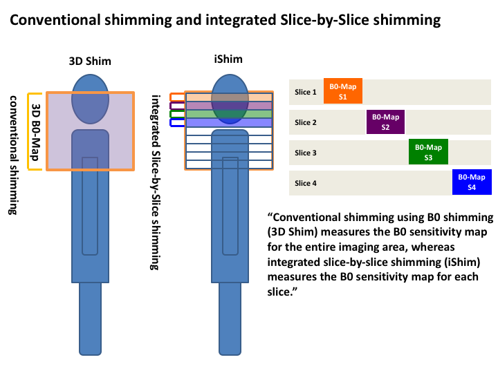
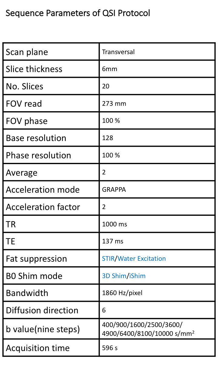
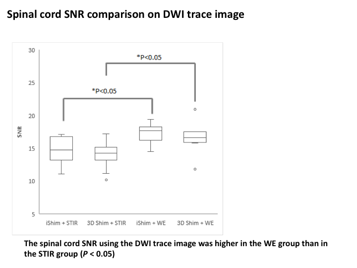
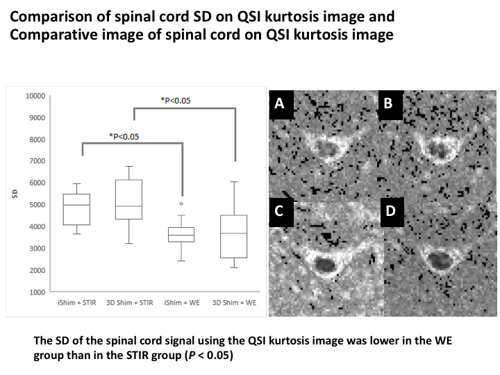
Figure 4 Left: Comparison of spinal cord SD on QSI kurtosis image
Right: Comparative image of spinal cord on QSI kurtosis imageA: iShim + STIR; B: 3D shim + STIR; C: iShim + WE; D: 3D shim + WE
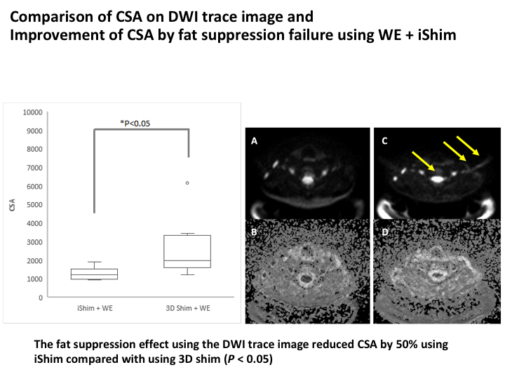
Figure 5 Left: Comparison of CSA on DWI trace image
Right: Improvement of CSA by fat suppression failure using WE + iShim A: DWI trace image iShim + WE; B: QSI kurtosis image iShim + WE; C: DWI trace image 3D shim + WE; D: QSI kurtosis image 3D shim + WE