1624
Progress in the use of SQUASHER for Diffusion weighted imaging1Center for Magnetic Resonance Research, University of Minnesota, Minneapolis, MN, United States, 2Department of Electrical and Computer Engineering, University of Minnesota, Minneapolis, MN, United States
Synopsis
The applicability of SQUASHER to EPI, along with a kz-dependent reconstruction approach for highly-accelerated 3D segmented EPI in dMRI
Introduction
High-resolution diffusion MRI (dMRI) remains challenging due to low SNR and long scan times. Several approaches have been proposed for high-resolution dMRI, including segmented acquisitions (1) and echo volumetric imaging (2). However, the increased scan time in the former, and the increased T2 blurring and signal loss in the latter have hindered their utility. An alternative line of work aims at imaging thicker slices multiple times, and resolving them at a higher resolution in the reconstruction (3,4). The original SLIDER approach (3) images slices with sub-voxel spatial shifts, which was shown to have unfavorable noise amplification (5). Subsequently, this was replaced with gSLIDER (4), which uses different RF excitation profiles to image the same slice/slab to improve the conditioning of the reconstruction. In order to avoid the sharp transitions of the RF profiles in gSLIDER, an alternative 3D encoding method called SQUASHER (Slice quadratic phase with HSn encoding and reconstruction) was also proposed (6), in which a quadratic phase is imposed on the slab, which spreads its k-space spectrum. In this work, we investigate the applicability of SQUASHER to EPI, along with a kz-dependent reconstruction approach for highly-accelerated 3D segmented EPI in dMRI.Methods
SQUASHER 3D-EPI: A 3D-EPI acquisition with slice and phase-encoding segmentation using frequency swept pulses for excitation and refocusing(4) was implemented to image thin slabs of less than 10 slices. A single navigator was used across all segments. Diffusion weighting was achieved using the spoiler gradients. Fast imaging by sub-sampling in the kz direction was evaluated with retrospective undersampling.
Reconstruction: Navigator correction was performed with linear phase-difference estimated from the navigator, and applied channel-specific. To evaluate the robustness of sub-sampling in kz direction, both a conventional 3D-GRAPPA algorithm5 and a kz-dependent 3D-GRAPPA algorithm(7) were implemented. The latter builds on an earlier modified GRAPPA approach(6), and utilizes a region-specific 3D kernel for different slice encoding locations.
Phantom imaging, A single-shot partition segmented diffusion weighted SE-EPI with TE/TR=143/4000ms, 1.5mm3 isotropic resolution, 8 slices/slab, 25% slab oversampling was used. This was used to compare the spectrum of the SQUASHER 3D-EPI against a standard implementation with a SINC RF-pulse. The SQUASHER data was retrospectively sub-sampled at rate 4×2 (ky × kz), and reconstructed using single-kernel and kz-dependent 3D-GRAPPA approaches.
In-vivo imaging was performed on a 3T Prisma, equipped with a 32 channel receiver coil. For GRAPPA kernel estimation, an ACS reference scan with 1 segment in the phase-encoding direction was acquired with TE/TR=129/800ms, 1.5x1.5x1mm3 resolution, 8 slices/slab, 25% slab oversampling, matrix size=128x128x10 and matched RF to the accelerated acquisition. For accelerated acquisitions, phase-encoding segmentation (two) was used, with TE/TR=91/800ms, FOV and resolution matching the reference scan. These acquisitions were repeated both with and without diffusion weighting.
Results
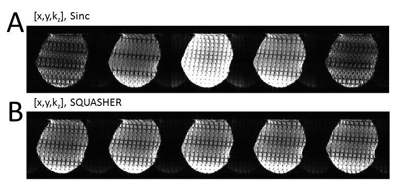
Fig. 1 shows the signal energy of the acquisitions using SQUASHER and SINC encoding pulses, in the x-y-kz plane following a root-sum-squares combination across coils. SINC encoding exhibits a fast decay in energy in the kz direction, which hinders segmentation due to the low SNR in the high-frequency components. On the other hand, SQUASHER exhibits a slow decay in the kz direction, which may be favorable for slice segmentation correction.
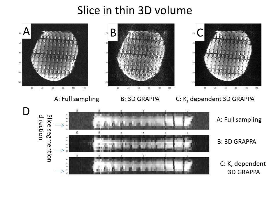
Fig. 2 depicts a phantom reconstruction with 4×2 undersampling. Single-kernel reconstruction (b) suffers from residual aliasing and signal variation, whereas the kz-dependent kernels (b) enable a distortion-free image at this high acceleration rate. The signal variation in a reformatted view (d) shows the superiority of the kz-dependent kernels in maintaining the signal variations across slices in this thin slab.
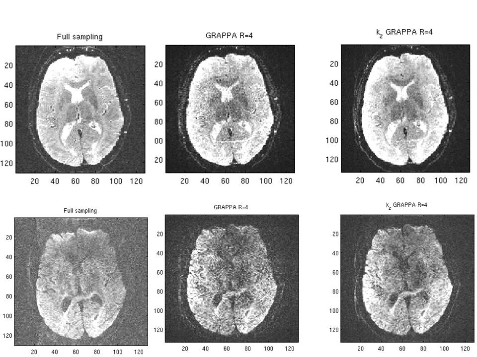
Fig. 3 shows the application of these techniques to human brain imaging for a single slice from a 3D acquisition at 4-fold ky acceleration, with 2 different diffusion weightings, and with 1mm through plane resolution and 1.5x1.5mm in-plane resolution.
Discussion
The use of SQUASHER and the kz-dependent GRAPPA algorithm allows for a higher SNR acquisition and better reconstruction. These provide a flexible, scalable approach for high resolution imaging with 3D-acquisitions of thin slabs.
For 3D thin-slab high resolution imaging, one of the challenges for the slice-segmentation is that the acquired kz slices have low signal. The low signal was previously addressed with the gSLIDER technique (4) where a Hadamard type encoding with n different RF pulses was used with n slices having coherent RF phase for increased signal. In contrast, SQUASHER requires a single RF encoding shape, while maintaining the conventional Fourier slice and phase encoding, but instead modifies the properties of the k-space signal for better segmentation.
In this study, we have shown the feasibility of combining SQUASHER and the kz-dependent GRAPPA for accelerated 3D EPI diffusion weighted imaging. Higher-resolution acquisitions and combination with SMS will be explored in future studies for faster whole brain coverage.
Acknowledgements
Grant support: NIH P41 EB015894, NIH U01 EB025144 and NSF CAREER CCF-1651825.References
1. Frost R, Miller KL, Tijssen RH, Porter DA, Jezzard P. 3D multi-slab diffusion-weighted readout-segmented EPI with real-time cardiac-reordered K-space acquisition. Magn Reson Med 2014;72(6):1565-1579.
2. Jeong EK, Kim SE, Parker DL. High-resolution diffusion-weighted 3D MRI, using diffusion-weighted driven-equilibrium (DW-DE) and multishot segmented 3D-SSFP without navigator echoes. Magn Reson Med 2003;50(4):821-829.
3. Setsompop KB, Berkin Nummenmaa, Aapo Fan, Qiuyun Cauley, Stephen Huang, Susie Yi Chatnuntawech, Itthi Rathi, Yogesh Witzel, Thomas Wald, Lawrence L. . SLIce Dithered Enhanced Resolution Simultaneous MultiSlice (SLIDER-SMS) for High Resolution (700 Um) Diffusion Imaging of the Human Brain. ISMRM 2015.
4. Setsompop K, Fan Q, Stockmann J, Bilgic B, Huang S, Cauley SF, Nummenmaa A, Wang F, Rathi Y, Witzel T, Wald LL. High-resolution in vivo diffusion imaging of the human brain with generalized slice dithered enhanced resolution: Simultaneous multislice (gSlider-SMS). Magn Reson Med 2017.
5. Moeller SS, Sebastian Akcakaya, Mehmet Noise Amplification Vs. Resolution Tradeoff in the SLIDER Technique. ISMRM 2016:3261.
6. Moeller SW, Xiaoping Harel, Noam Garwood, Mike Akcakaya, Mehmet SQUASHER: Slice Quadratic Phase with HSn Encoding and Reconstruction. ISMRM 2017:1522.
7. Steen Moeller SR, Essa Yacoub, Mehmet Akcakaya. Improved 3D parallel imaging with localized estimation of 3D GRAPPA kernels. ISMRM 2018.
Figures