1623
Low b-values and limited diffusion directions introduce bias in FA and MD that increases with decreasing voxel volumes.1Department of Human Biology, University of Cape Town, Cape Town, South Africa, 2Cape Universities Body Imaging Centre (CUBIC), University of Cape Town, Cape Town, South Africa
Synopsis
Due to ECG triggering and breath hold techniques required to compensate for motion of the beating heart and respiration, acquisition times for cardiac diffusion tensor imaging (DTI) are limited. As such, lower b-values and fewer diffusion directions are typically used, together with larger slice thicknesses. This study aims to assess the impact of these changes on fractional anisotropy (FA) and mean diffusivity (MD) in a pineapple phantom. Smaller voxels were found to be more sensitive to changes in b-values and number of diffusion directions.
Introduction
Diffusion tensor imaging (DTI) offers a non-invasive method of studying the microstructural organisation of tissue1. Typically used for brain imaging, recent applications have demonstrated the potential use of DTI for imaging other organs, most notably the heart2. Synchronization with the beating heart and breath hold techniques to correct for respiratory motion limit acquisition times, necessitating low b-values and fewer diffusion directions. To cover the entire heart in fewer acquisition, thicker slices are also used, leading to anisotropic voxels. Here we examine the impact of these changes on fractional anisotropy (FA) and mean diffusivity (MD). We hypothesized that anisotropic voxels would introduce bias in FA and MD values.Methods
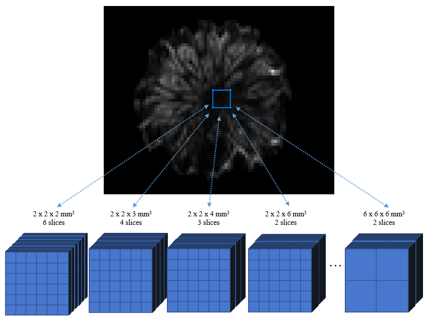
DTI data of a pineapple phantom was acquired using a twice-refocused spin echo DTI pulse sequence3 on a 3T Skyra MRI (Siemens, Erlangen, Germany). A 15-channel knee coil was used to obtain a higher SNR. To examine the effect of voxel dimensions, acquisitions were repeated for in-plane voxel sizes 2.0 x 2.0 mm2, 3.0 x 3.0 mm2, 4.0 x 4.0 mm2, and 6.0 x 6.0 mm2, each for slice thicknesses 2.0 mm, 3.0 mm, 4.0 mm and 6.0 mm. To imitate the conditions used in cardiac and brain DTI, each acquisition was repeated for b-values 300 and 1 000 s/mm2, and for 6 and 30 diffusion directions, respectively. The remaining acquisition parameters were as follows: TR = 1200 ms, TE = 100 ms, FOV = 384 x 384 mm2, flip angle = 90°, and bandwidth = 1565 Hz/pixel.
Diffusion tensor estimations were performed using the weighted least linear squares algorithm in ExploreDTI4. DTI parameters were computed in a fixed region of interest (ROI, 12 x 12 x 12 mm3) in the core of the pineapple. The average FA and MD across the ROI were compared for different voxel dimensions (Fig. 1), and for different b-values and number of diffusion directions.
Results
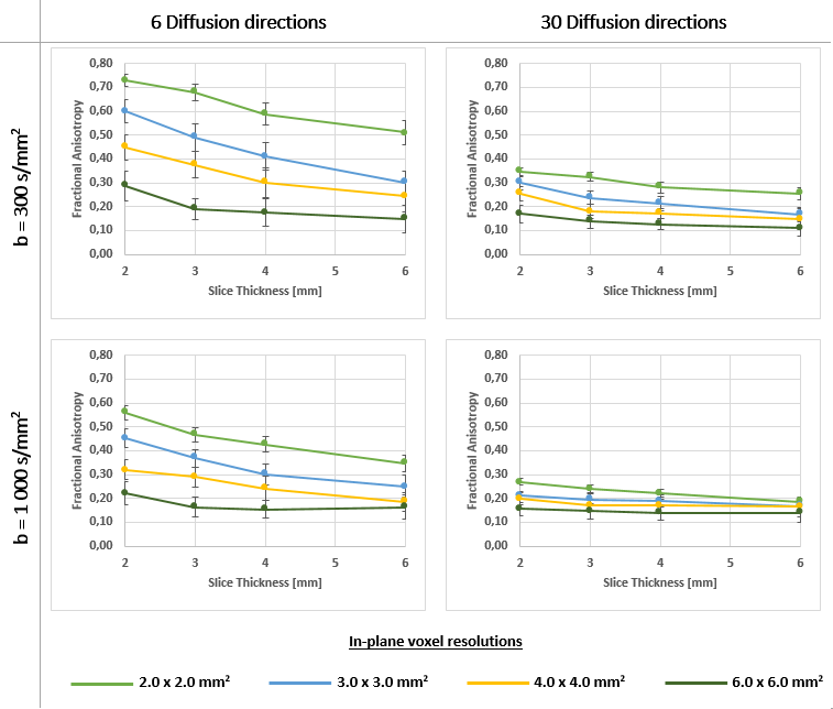
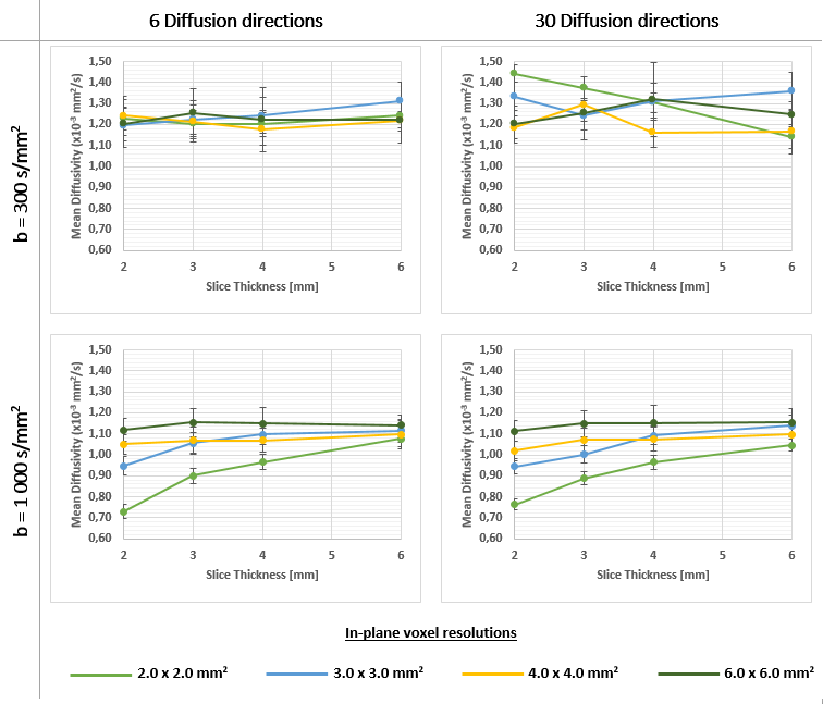
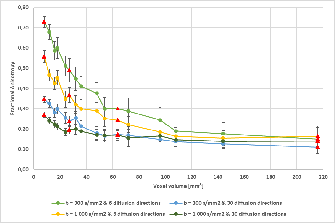
Figures 2 and 3 show the average FA and MD values, respectively, across the ROI for different spatial resolutions, and for low and high b-values (rows) and 6 or 30 diffusion directions (columns). FA and MD values for b-value 1 000 s/mm2 and 30 directions were used as baseline. Reducing the number of diffusion directions and the b-value independently and combined resulted in FA increases, with the largest increases occurring in the smallest voxels. Similarly, reducing the b-value caused MD increases that were greatest in the smallest voxels. In contrast, MD values were less affected by changes in the number of diffusion directions. Figure 4 shows that FA values increase with decreasing voxel volumes for all b-values and diffusion directions. As such, the observed FA increases are not attributable to in-plane resolution, slice thickness or voxel anisotropy.Discussion
These findings show that FA and MD are greatly impacted by voxel volume, and that the effect of voxel volume is exaggerated at low b-values and for limited diffusion directions. These results indicate that larger voxel dimensions may be more appropriate in cardiac DTI applications where b-values and the number of diffusion directions are limited by timing constraints resulting from ECG gating and breath hold techniques. Contrary to our hypothesis, neither FA nor MD were affected by voxel anisotropy.Conclusion
In applications requiring low b-values and fewer diffusion directions, larger voxels may yield more accurate FA and MD estimates.Acknowledgements
- South African Research Chairs Initiative - NRF SARC06052016A
- The Mandela Rhodes Foundation
References
1. D. K. Jones and A. Leemans, “Diffusion Tensor Imaging,” Methods Mol. Biol., vol. 711, pp. 127–144, 2011.
2. C. Mekkaoui, T. G. Reese, M. P. Jackowski, H. Bhat, and D. E. Sosnovik, “Diffusion MRI in the heart,” NMR Biomed., no. September, 2015.
3. T. G. Reese, O. Heid, R. M. Weisskoff, and V. J. Wedeen, “Reduction of eddy-current-induced distortion in diffusion MRI using a twice-refocused spin echo,” Magn. Reson. Med., vol. 49, no. 1, pp. 177–182, 2003.
4. A. Leemans, B. Jeurissen, J. Sijbers, and D. . Jones, “ExploreDTI: a graphical toolbox for processing, analyzing , and visualizing diffusion MR data,” in ISMRM 17th Scientific Meeting & Exhibition, 2009, vol. 245, no. 2, p. 3537.
Figures