1620
Comparison of different diffusion MRI acquisition protocols by tracking callosal motor pathways with deterministic and probabilistic fiber tracking algorithms1Center for MRI Research, Peking University, Beijing, China
Synopsis
High angular resolution diffusion MRI (HARDI), the most widely used method in in-vivo brain imaging experiments to delineate white matter pathways, has been found sufficient for resolving 2-way fiber crossings but unstable for detecting 3-way fiber crossings. Therefore, if more sensitive and accurate tractography is wanted, researchers need to use high b-value with multi-shell q-ball models, which can be time-consuming. In this study, we compared 3 diffusion MRI acquisition protocols by tracking callosal connections between motor areas with both probabilistic and deterministic fiber tracking algorithms and provided a new scheme for the future diffusion MRI experiment.
Purpose
Diffusion MRI tractography is widely used in in-vivo brain imaging experiments to delineate white matter pathways in human brain. Nowadays, it is common practice to use high angular resolution diffusion MRI (HARDI) with single-shell gradient table, and the b-value is typically around 1000 s/mm2. Previous research have found that the diffusion tensor MRI (DTI) data acquired with this conventional protocol is sufficient for resolving 2-way fiber crossings but unstable for detecting 3-way fiber crossings1. Therefore, for some occasions, high b-value with multi-shell DWI protocol is needed for a more sensitive and accurate tractography2,3, which can be time-consuming. For example, the DWI protocol of Human Connectome Life Span Project (LS) is designed in q-ball models, and the data is acquired with 2 gradient tables (UM_98dir and UM_99dir), and each direction is acquired with phase-encode A>>P and P>>A, taking more than 20 minutes, which is too long for common diffusion MRI experiments to be widely used.
In this abstract, we attempted a new acquisition scheme, in which we used the same gradient table as LS, but data for 2 gradient tables are collected in opposite phase-encoding directions, respectively. We compared both deterministic and probabilistic fiber tracking results of conventional HARDI protocol (Protocol 1), LS DWI protocol (Protocol 2) and the newly proposed acquisition scheme (Protocol 3). In this way, we hope to find a more time-efficient acquisition protocol with relatively higher image quality and put forward a new option for future diffusion MRI experiments.
Methods
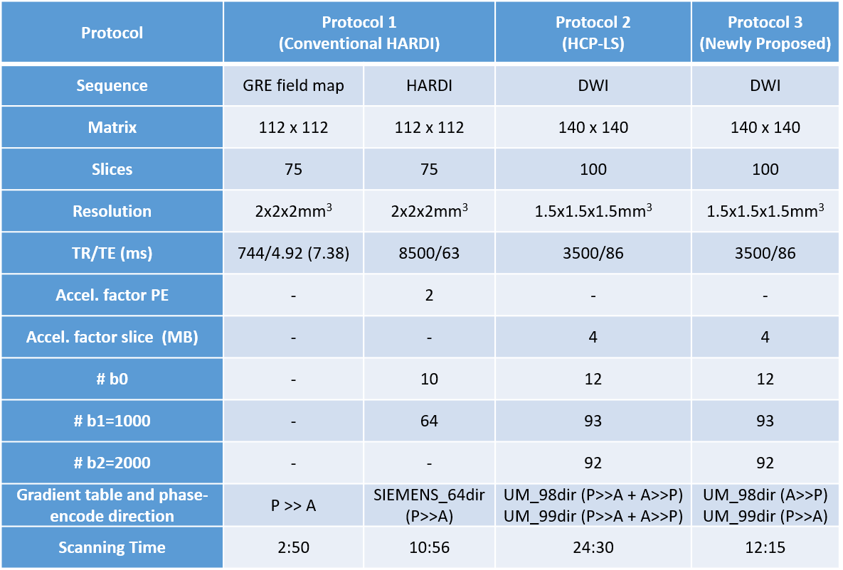
Data Acquisition: One right-handed healthy volunteer was scanned in a 3T MRI system (Siemens Magnetom Prisma). T1-weighted 3D structural images were obtained using MPRAGE sequence (TR/TE/FA=2400s/2.22ms/8°, 0.8x0.8x0.8mm3). And then, whole brain diffusion images were acquired with 3 protocols described above. Furthermore, gradient-echo sequence(GRE) with two different TEs was scanned using the conventional field-mapping method for distortion correction of Protocol 1. The detailed scan parameters were listed in Table 1.
Data Processing: All the dMRI data were preprocessed by FSL in individual diffusion space. Data from Protocols 2&3 were processed with the TOPUP method, and data from Protocol 1 was processed using the conventional GRE-based field-mapping method. ROIs of 3 motor areas (face areas, hand areas, foot areas) were drawn bilaterally in individual diffusion space based on the anatomical atlas. The fiber tracts of callosal motor pathways were delineated with DTI and q-ball models using deterministic and probabilistic fiber tracking algorithms. Probabilistic fiber tracking is calculated by FSL bedpostx, and deterministic fiber tracking is calculated by DiffusionKit (brainnetome dMRI Toolkit).
Results and Discussion
As is shown in table 1, Protocol 1 collected the conventional field map and the HARDI sequence, which takes more than 13 minutes in total. Protocol 2 collected both A>>P and P>>A phase-encode directions for 2 gradient tables, taking around 25 minutes. Protocol 3 collected A>>P phase-encode direction for the first gradient table (UM_98dir) and P>>A phase-encode direction for the other gradient table (UM_99dir), which just takes around 12 minutes, less than Protocol 1 in time-consuming.
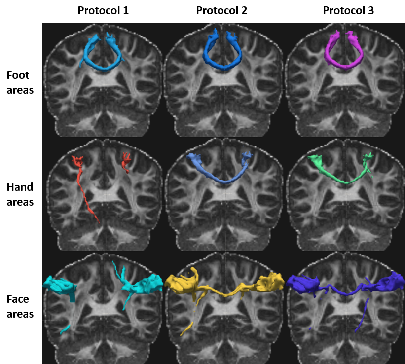
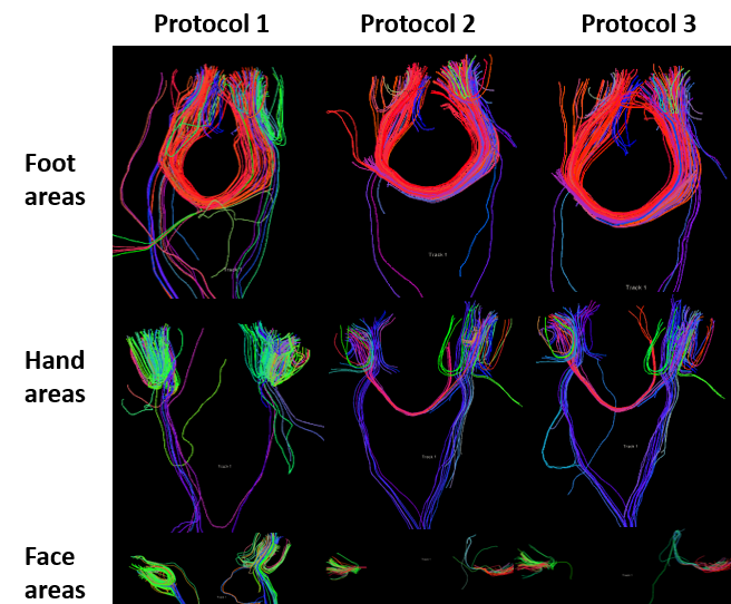
The probabilistic fiber tracking results of DWI data from the 3 protocols were shown in Fig.1. For one motor area, the lower thresholds in tractography results were set the same among different protocols for comparisons. Base on the tractography results from both probabilistic (Fig.1) and deterministic (Fig.2) fiber tracking algorithms, it is obvious that the newly proposed method (Protocol 3) generated better results than the conventional HARDI protocol, and it also obtained similar fiber tracts as the LS protocol.
Conclusion
In this study, we compared 3 different dMRI acquisition protocols by tracking callosal connections between motor areas, including foot areas, hand areas and face areas. These preliminary results showed that the newly proposed protocol (Protocol 3) has potentials in getting similar tractography results as the LS DWI protocol (Protocol 2) but only takes half the time. This comparative study provides a new scheme for the future dMRI experiment.Acknowledgements
I want to take this chance to thanks to my advisor Jia-Hong Gao, a professor of School of physics in Peking University. In the process of composing this paper, he gives me many academic and constructive advices, and helps me to correct my paper. Except these, he also gave me the opportunity to expand my practice.References
1. Sotiropoulos, S.N., et al., Advances in diffusion MRI acquisition and processing in the Human Connectome Project. Neuroimage 2013; 80: 125-43.
2. Tuch, D.S., et al., High angular resolution diffusion imaging reveals intravoxel white matter fiber heterogeneity. Magn Reson Med. 2002; 48(4): 577-82.
3. Jbabdi, S., et al., Model-based analysis of multishell diffusion MR data for tractography: how to get over fitting problems. Magn Reson Med. 2012; 68(6): 1846-55.
Figures