1619
Evaluation of Monopolar Diffusion-Prepared TSE for Diffusion Imaging1Interdisciplinary Institute of Neuroscience and Technology, Qiushi Academy for Advanced Studies, Zhejiang University, Hangzhou, China, 2College of Biomedical Engineering & Instrument Science, Zhejiang University, Hangzhou, China, 3Center for Magnetic Resonance Research, School of Medicine, University of Minnesota, Minneapolis, MN, United States, 4Key Laboratory for Biomedical Engineering of Ministry of Education, Zhejiang University, Hangzhou, China
Synopsis
EPI-based diffusion imaging methods are dominantly used, but suffer from susceptibility associated distortion and signal loss, making it challenging to achieve high-quality high-resolution diffusion imaging results. To overcome these challenges, we implemented monopolar diffusion preparation module for TSE sequence (DP-TSE) and evaluated the performance in comparison to readout segmented multi-shot echo planner (RESOLVE) sequence for diffusion weighted imaging (DWI). Our study results suggest that Diffusion-Prepared TSE is a promising alternative for distortion-free, high-resolution diffusion imaging with superior diffusion SNR.
Purpose
Diffusion MRI, an essential method for investigation of tissue microstructure, has been widely used in both neuroscience research and clinical applications, such as human connectome project and white matter diseases studies.1-3 However, the most commonly used image readout for diffusion imaging is echo planar imaging (EPI), which suffers from susceptibility associated image distortion and signal loss, making it challenging to achieve high quality high-resolution diffusion imaging. In contrast, turbo-spin echo (TSE) image, less sensitive to susceptibility and capable to provide superior signal-to-noise ratio (SNR) for diffusion images, can be a good alternative image readout. We implemented and evaluated a diffusion imaging method with the TSE as the image readout, called Diffusion-Prepared TSE (or DP-TSE), and the results from our initial studies are reported here.Methods
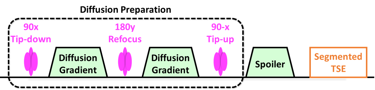
Studies were performed on a honey-dew melon (phantom) and several healthy human subjects on a 3T clinical MR scanner (Magnetom Prisma, Siemens Healthcare, Erlangen, Germany) under an IRB approved protocol with written informed consent. The body coil was used for RF transmission and a 32-channel phased array head coil for signal reception. The sequence diagram of the implemented imaging method is presented in Figure 1. A monopolar, instead of a bipolar, diffusion preparation module using a pair of Stejskal and Tanner diffusion gradients4 was used to reduce diffusion preparation time and minimize the signal loss due to T2 decay. Two optimized spoiler gradients were applied around the preparation module to eliminate any potential residual signal.
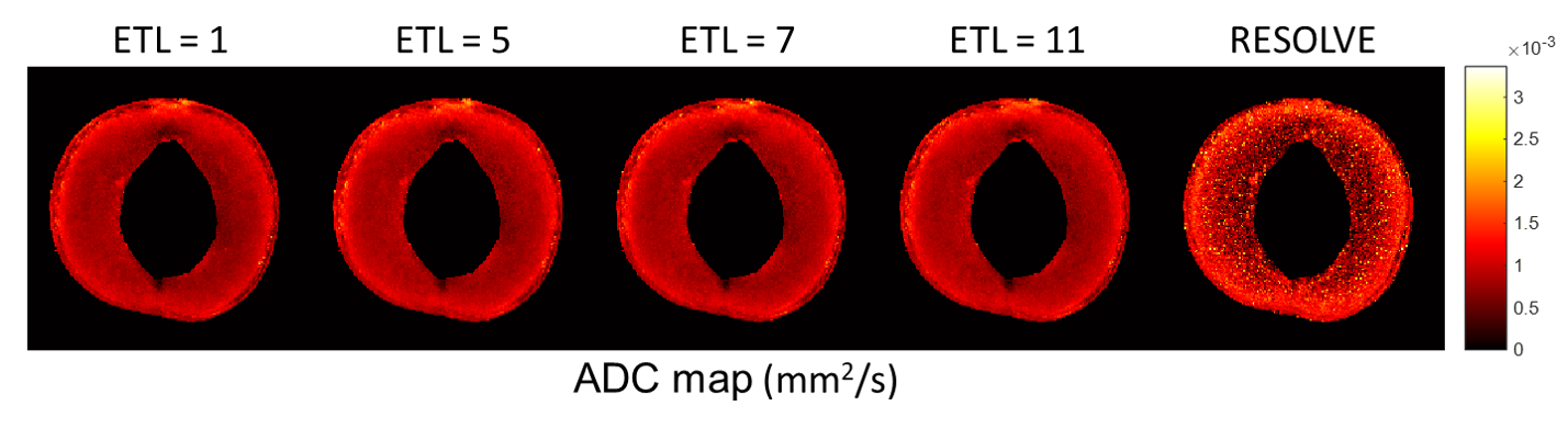
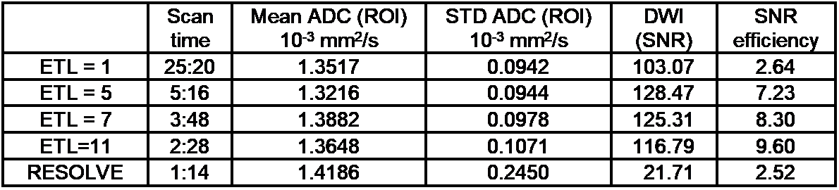
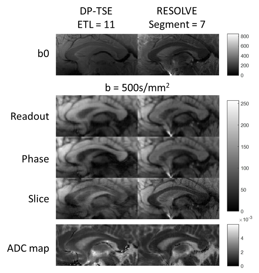
To assess the accuracy of the implemented method and investigate the balance between imaging quality and acquisition time, diffusion imaging studies with the honey-dew melon were first performed using a series of b-values along with varied echo train lengths (ETLs): FOV = 192 x 192 mm2; matrix size = 192 x 192; in-plain resolution = 1.0 x 1.0 mm2; slice thickness = 3 mm; TE = 10 ms; TR = 2000 ms; ETL = 1, 5, 7, 11; and b-value = 500, 1500 s/mm2. To further evaluate the implemented method, especially its robustness for in vivo imaging, diffusion studies of the corpus callosum were performed with a series of volunteers using the following parameters: FOV = 240 x 240 mm2; matrix size = 192 x 192; in-plain resolution = 1.25 x 1.25 mm2; slice thickness = 3 mm; TE = 10 ms; TR = 1500 ms; ETL = 11; b-value = 500, 1000 s/mm2. For comparison purpose, in vivo multi-shot RESOLVE diffusion imaging was also performed using matched parameters with 7 readout segments. Post-processing, including co-registration and reconstruction of apparent diffusion coefficient (ADC) maps, were performed in MATLAB
Results and Discussions
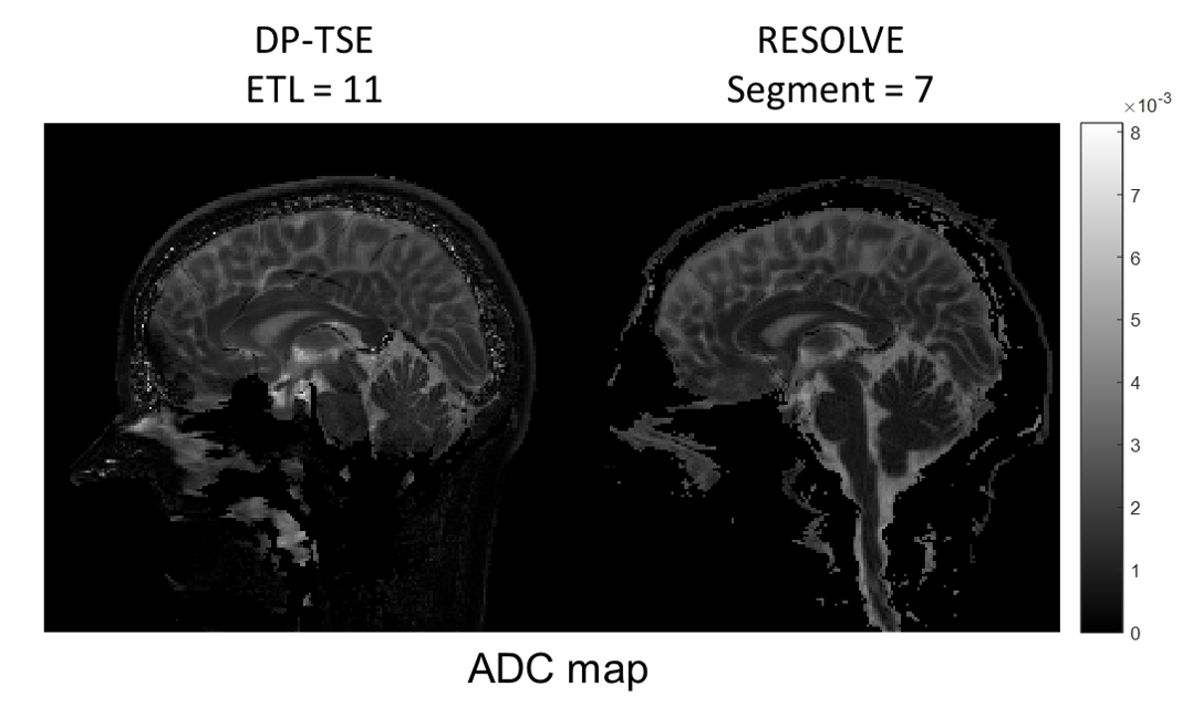
ADC map comparisons between DP-TSE and RESOLVE of melon are displayed in Figure 2, followed by a quantitative comparison for different DWI methods in scan time, mean/std ADC value, SNR and SNR efficiency summarized in Table 1. Diffusion-weighted images of corpus callosum and whole brain ADC map comparisons between DP-TSE and RESOLVE are shown in Figure 3 and 4.
The study results indicated that non-distorted, high SNR and accurate diffusion weighted images can be achieved by diffusion-prepared TSE sequence. ADC values were consistent with varied ETL numbers in DP-TSE DWI measurement. Although TSE-based DWI sequences5,6 still consumes more time than RESOLVE sequence, the SNR efficiency of DP-TSE is generally higher than 7-shot segmented RESOLVE. The diffusion signal higher intensity and SNR of DP-TSE may potentially help push the resolution of diffusion imaging to be higher.
In the human study, the ADC results of corpus callosum were comparable between DP-TSE and RESOLVE measurements. Our in vivo study results further confirmed that the DP-TSE method could provide ADC values comparable to those from RESOLVE and in literature7 (Figure 3), but suffers less from susceptibility distortion (Figure 4). The limitation of DP-TSE is the potential sensitivity to motion, which may be addressed by motion insensitive trajectory such as PROPELLER or radial readout.
Conclusions
DP-TSE is a promising alternative for distortion-free, high-resolution diffusion imaging with superior diffusion image SNR.Acknowledgements
P41 EB015894, P30 NS076408 and Human Connectome Projects (U01 MH109589 and U01 AG052564), and UL1TR000114. This research work is also supported by the University of Minnesota Foundation. The content is solely the responsibility of the authors and does not necessarily represent the official views of the National Institutes of Health.References
- Johansen-Berg H, Behrens TE, editors. Diffusion MRI: from quantitative measurement to in vivo neuroanatomy. Academic Press; 2013 Nov 4.
- Vu AT, Auerbach E, Lenglet C, Moeller S, Sotiropoulos SN, Jbabdi S, Andersson J, Yacoub E, Ugurbil K. High resolution whole brain diffusion imaging at 7T for the Human Connectome Project. Neuroimage. 2015 Nov 15; 122:318-331.
- Song SK, Yoshino J, Le TQ, Lin SJ, Sun SW, Cross AH, Armstrong RC. Demyelination increases radial diffusivity in corpus callosum of mouse brain. Neuroimage. 2005 May 15; 26(1):132-40.
- Stejskal EO, Tanner JE. Spin diffusion measurements: spin echoes in the presence of a time‐dependent field gradient. The journal of chemical physics. 1965 Jan 1; 42(1):288-292.
- Zhang Q, Coolen BF, Versluis MJ, Strijkers GJ, Nederveen AJ. Diffusion‐prepared stimulated‐echo turbo spin echo (DPsti‐TSE): An eddy current‐insensitive sequence for three‐dimensional high‐resolution and undistorted diffusion‐weighted imaging. NMR in Biomedicine. 2017 Jan 1.
- Pipe JG, Farthing VG, Forbes KP. Multishot diffusion‐weighted FSE using PROPELLER MRI. Magnetic resonance in medicine. 2002 Jan 1;47(1):42-52.
- Moon WJ, Provenzale JM, Sarikaya B, Ihn YK, Morlese J, Chen S, DeBellis MD. Diffusion-tensor imaging assessment of white matter maturation in childhood and adolescence. American Journal of Roentgenology. 2011 Sep; 197(3):704-712.
Figures