1611
Diffusion Weighted Signal Variation with Body Phantom1AIM Medical Imaging, Vancouver, BC, Canada
Synopsis
DWI body phantom development and signal to noise calculation based on RSNA QIBA protocol guidelines for identically configured MRI machines shows machine variability and resultant ADC calculation error propagation.
Introduction
Diffusion weighted imaging (DWI) is a commonly used MRI sequence that assesses for water mobility between two motion probing gradients. The applied strength of these temporary motion probing gradients [b-values] results in changes to the water signal: as b-values increase, typically lesions that restrict water mobility due to increased nuclear cytoplasmic ratio, or cell density retain signal, while the adjacent normal tissue background signal is lost. These differences in signal at different b-values can be used to calculate a slope which represents the apparent diffusion coefficient [ADC], which is purported to be a quantifiable reproducible machine independent value. However, the impact on signal to noise with multiple b-values, or at various geographic cross-sectional locations within an axial slice has not been previously measured.Methods
A homogeneous material phantom was made using QIBA materials [ref 1] with a standard 32 inch body circumference with a 5cm thickness. A second simulated human body phantom with variably restricted material and gas components was also made. Multiple 1.5T machines from the same vendor [Siemens Aera] using the same coil set, software version and DWI sequence parameters (b0,500,900,2000 with advanced shim, 50% oversampling, 2averages, parallel factor=2) at isocenter were used for scanning the DWI homogeneous material and simulated human body phantoms. For each of the phantoms, two scans were obtained sequentially without moving the phantom, and multipass signal to noise (SNR) was calculated for DWI according to ACR-NEMA procedure guidelines utilized by QIBA [ref 1].Results
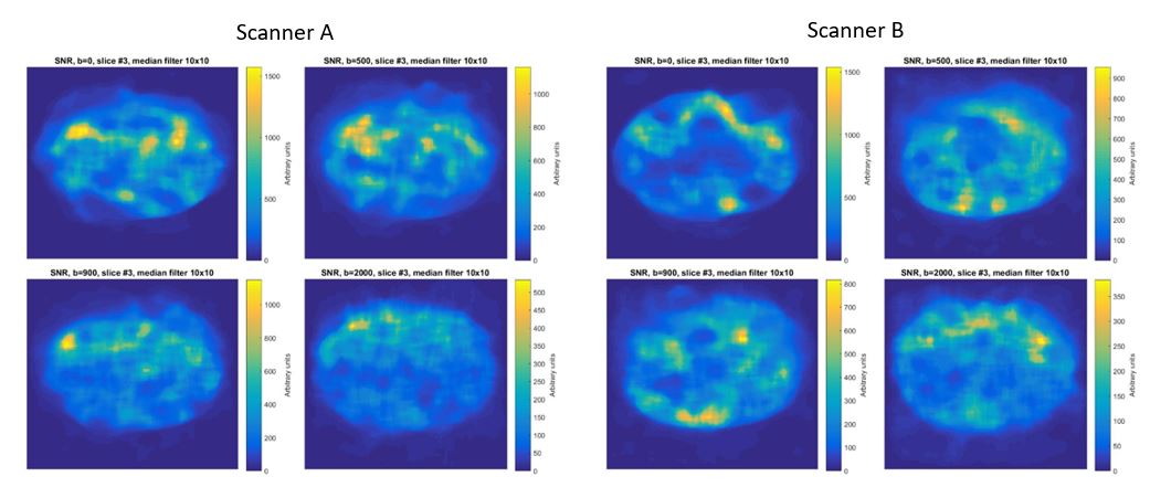
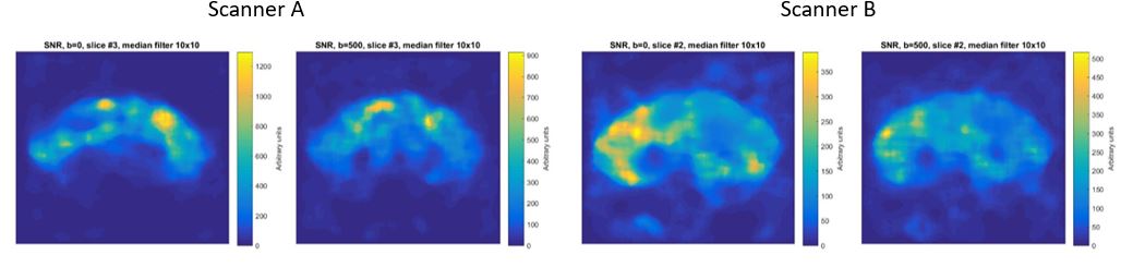
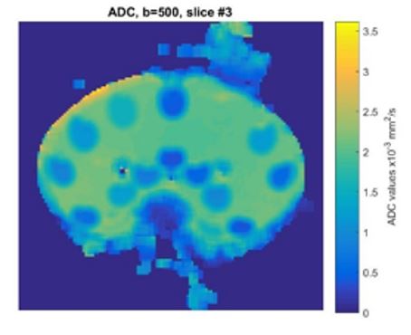
Cross sectional images at isocenter for b-values of 0, 500, 900 and 2000 are shown in Figure 1 for the homogeneous phantom, where the signal is measured in arbitrary units for 2 identically configured 1.5T machines. Figure 2 shows the cross sectional SNR profile for the simulated human body phantom. Figure 3 shows the calculated ADC value across the axial isocenter profile for b-value of 500 and 0 using the simulated body phantom.Discussion
With a homogeneous material phantom, the SNR at multiple b-values for a single scanner is inconsistent demonstrating no specific pattern to the degree of variability; either between geographic location [i.e. coil proximity], or with increased b-value motion probing gradients.
Additionally between 2 identically configured machines, the SNR at the same geographic location and at the same b-values is also inconsistent. As the underlying b-value SNR is critical for calculation of ADC, this heterogeneity in geographic signal will propagate into the calculation, limiting both the accuracy and precision of the ADC metric.
The impact of b-value SNR variability is exacerbated with the human body simulated phantom, which demonstrates greater signal to noise variability between identical MRI machines as shown in Figure 2. The impact of this SNR variability at geographic locations and resultant signal distortion is demonstrated in the ADC calculation of Scanner A calculated from b 0 and 500. The ADC values shown in Figure 3 also demonstrate warping and susceptibility artifact that arises from the inherent errors in b-value. These errors demonstrate typical ghosting effects which clinically were thought to be due to patient motion or susceptibility at gas/tissue interfaces.
The impact of hardware variability versus software variability cannot be ascertained by this current study, as only the SNR of the DICOM images were assessed.
Conclusion
DWI continues to grow in clinical relevance, particularly in the field of body oncologic studies. However the geographic heterogeneity in signal for body imaging and resultant propagation into ADC values as well as lack of MRI machine interchangeability at 1.5T demonstrated with this phantom-based study will hinder setting reliable numeric ADC cutoffs for tissue identification and classification. The SNR variability at higher static and gradient field strengths needs to be measured as susceptibility effects become more pronounced with increasing static field strengths.Acknowledgements
Drs. Michael Boss, Tom Chenevert and Dariya Malyarenko as well as the QIBA DWI committee.References
1. http://qibawiki.rsna.org/index.php/Perfusion,_Diffusion_and_Flow-MRI_Biomarker_CtteFigures