1600
Intra- and inter-subject variability of diffusivity by DTI and DKI: An small animal study on 7T1Biomedical Engineering and Environmental Science, National Tsing Hua University, Hsinchu, Taiwan, 2Department of Biomedical Imaging and Radiological Science, China Medical University, Taichung, Taiwan, 3Linkou Chang Gung Memorial Hospital, Taoyuan, Taiwan
Synopsis
Diffusivity can be acquired by both DTI and DKI model on the same set of images. To investigate intra- and inter-subject variability of DKI and DTI derived diffusivities, five Sprague- Dawley rats were scanned on a 7T small animal scanner. In intra-subject variability test, lower coefficients of variation are found on DKI derived parameters. In inter-subject analysis, higher values were estimated by DKI in mean diffusivity, axial diffusivity, and radial diffusivity. The CNR between white matter and gray matter of these parameters are also better with DKI. However, the CNR of FA is higher with DTI than with DKI
Introduction
Several studies have shown that Diffusion kurtosis imaging (DKI) performs better than DTI on neural tissue characterization and detecting alterations 1,2,3, especially in the gray matter. Instead of fitting one diffusivity parameter from monoexponential decay, both diffusivity and kurtosis parameters are estimated with a in a sufficient b-value range to obtain reliable fitting for Dapp and Kapp in the kurtosis model. However, introducing an additional parameter may influence the accuracy and precision in fittng procedure. In this study, we compared the variability of diffusivity from single exponential decay and kurtosis model. In order to acquire consistent diffusion weighted images, normal brains of Sprague- Dawley (SD) rats were scanned on a 7T scanner.Materials and Methods
SD rat (N= 5) were scanned on 7T MR Bruker Clinscan MRI. Animals were anesthetized with with 1.5% isoflurane mixed with oxygen. DWI images of Thirty gradient directions and 6 b-values were acquired (0, 500, 1000, 1500, 2000, and 2500 s/mm2) with TR/TE= 3000/32 ms, slice thickness= 1mm, FOV= 30 mm, matrix size= 92x 92. All data were fitted by DTI (i.e. single exponential decay) and DKI model. DKI parameters were fitted by nonlinear regression (lsqcurvefit function in MATLAB) with pre-fitting monoexponential decay D as initial guess value. Diffusional parameters such as mean diffusivity (MD), fractional anisotropy (FA), and axial and radical diffusivity (AD, RD) were calculated. Multi-slice regions of interest (ROIs) were manually delineated on FA maps. White matter (WM) ROIs include corpus callosum (CC) and external capsule (EC); gray matter (GM) ROIs include cerebral cortex (CT), and hippocampus (HP).Results
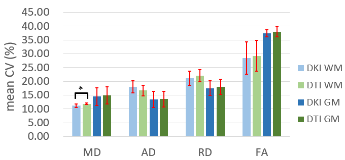
To investigate intra-subject variability, we first calculated coefficient of variation (CV) of MD, AD, RD, and FA within GM and WM of each rat, and then evaluated the average CV of 5 rats in Fig. 1. Except AD of WM, mean CVs of diffusivity with DKI model were lower than those with DTI model. The CV of MD in WM is significant lower in DKI model than that in DTI model (p<0.05).
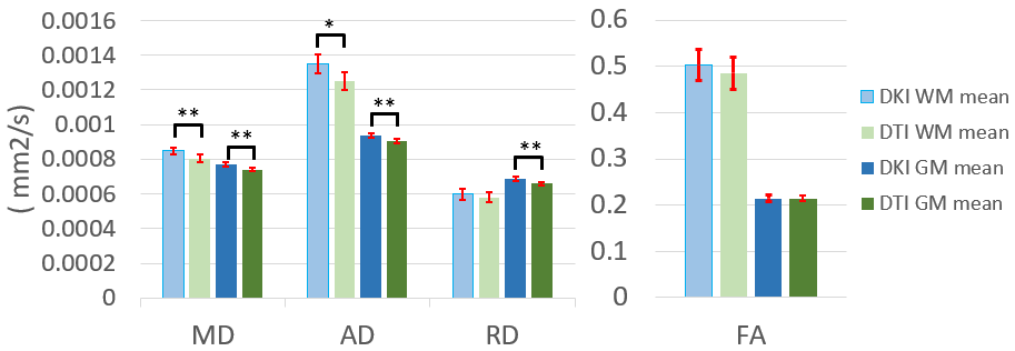
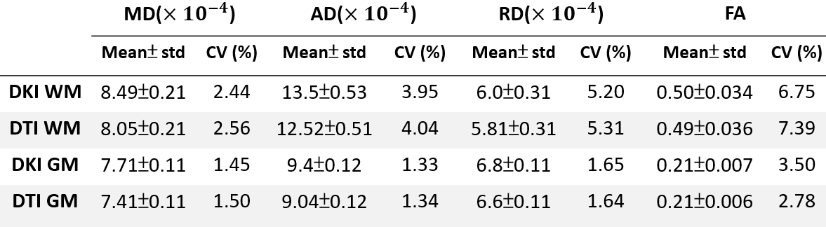
For inter-subject analysis, we show the average value and standard deviation of MD, AD, RD, and FA from 5 rats in Fig. 2. The CVs of inter-subject variation were showed in Table 1. The MD of WM, MD of GM, AD of WM, AD of GM, and RD of GM are significantly higher in DKI model than those in DTI model. The FAs of WM and GM are almost identical in two models. The CVs of inter-subject variation of each diffusivity parameter are very similar.
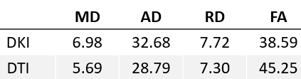
Furthermore, CNR of diffusivity between WM and GM were shown in table 2, which divided the difference between GM and WM by the standard deviation of GM.
Discussion
For intra-subject variability test, we assumed that tissue within WM and GM are uniform, CVs of DKI did not display higher variations in chosen ROIs, which demonstrated there are no decline in the precision of diffusivity when adding a Kapp in the nonlinear fitting procedure.
Inter-subject variability analysis revealed that there are significant differences in diffusivity mean values between DKI and DTI. However, CVs of diffusivities derived from DKI are lower than DTI, except for FA. DTI- and DKI- derived FA are nearly the same, due to the calculation of FA performed a normalized effect.
Furthermore, CNRs between WM and GM are higher in DKI-derived MD, AD, and RD than DTI-derived ones. According to literature 3, the sensitivity of MD, AD, and RD by DKI is higher than those by DTI. Because of DTI-derived directional diffusivities contain opposite and competing effects of diffusivity and kurtosis, the sensitivity of DTI-derived AD is diminished. However, due to the normalization effect in the calculation of FA, the CNR of FA by DKI is lower than that by DTI in our study.
Acknowledgements
No acknowledgement found.References
1. Hui ES et al., Towards better MR characterization of neural tissues using directional diffusion kurtosis analysis. Neuroimage 2008; 42: 122–133.
2. Cheung MM et al., Does diffusion kurtosis imaging lead to better neural tissue characterization? A rodent brain maturation study. Neuroimage 2009; 45:386–392.
3. Wu EX et al., MR diffusion kurtosis imaging for neural tissue characterization. NMR Biomed 2010; 23(7):836-48
Figures