1598
A non-Gaussian bi-exponential diffusion model with CUSP74 sampling for improved myocardial helix angle quantification and segmentation.1Anatomy and Medical Imaging, The University of Auckland, Auckland, New Zealand
Synopsis
The non-Gaussianity of diffusion at high b-value, leads to poor estimates of fast diffusion components when using diffusion models that assume Gaussian diffusion distributions. Including the diffusion kurtosis in a bi-exponential model allows better quantification of the partial volume effects when large b-values are used. This study investigates how this improved model can provide a better estimate of the helix angle in fixed heart specimens.
Introduction
Diffusion imaging of fixed cardiac specimens enables accurate quantification of myocardial fibre architecture. The presence of formalin in these specimens can adversely affect the calculation of cardiac diffusion metrics, such as fibre helix angle (HA). The tensor model used in diffusion tensor imaging (DTI) assumes a Gaussian distribution of diffusion. However, in tissues and at high b-values, the assumption of a Gaussian distribution does not hold1, 2, 3. Previous research4, 5 has shown that higher b-values are required to isolate the fast diffusion components originating in fluid. A non-Gaussian approximation accounting for the diffusion kurtosis6 provides better estimates of diffusion metrics at higher b-value compared to DTI7, 8. This study investigates the importance of including higher b-value and accounting for the non-Gaussian nature of diffusion in order to correct for partial volume effects and accurately determine cardiac diffusion metricsMethod
We scanned a formalin-fixed ex vivo sheep heart specimen with a monopolar spin echo sequence and a CUSP diffusion sampling method9. We used the “dmritool” package10 to form two uniformly distributed shells while keeping the selected points from all shells separated as far as possible. Two shells with 32 diffusion directions were respectively set at b=800s/mm2 and 800s/mm2<b<1500s/mm2, six directions at b=1700s/mm2 and for directions at b=2500s/mm2 to form CUSP74 sampling9. Acquisition parameters were: TR=2s, TE=56.88ms,$$$\delta=20.67\mu s$$$, $$$\triangle=27.09\mu s$$$, averages=1, Matrix=100x100, FOV=200x200, slice thickness=4mm and slices=12.
The diffusion distribution was determined using two bi-exponential models. The first assumed Gaussian distribution of diffusion in both the formalin and the tissue. The second assumed non-Gaussian diffusion in the tissue, accounting for the diffusion kurtosis, shown in Equation 1,
$$$\frac{S(b)}{S0}=(1-f)exp(-b\cdot D_{iso})+fexp(-b\cdot D_{tissue_{app}}+\frac{1}{6}b^{2}\cdot D_{tissue_{app}}^{2}\cdot K_{app})$$$
where S(b) and S0 are the signal with and without diffusion weighting, f is the volume fraction, $$$D_{iso}$$$ is the isotropic diffusion of the formalin, and $$$D_{tissue_{app}}$$$ and $$$K_{app}$$$ are the apparent diffusion coefficient of the tissue and the apparent diffusional kurtosis, respectively. Both bi-exponential models were compared to a mono-exponential model. $$$D_{iso}$$$ was determined using a mono-exponential fit in a region containing only formalin. The helix angle and volume fraction were calculated using each of the three models, in a manually selected region of interest around the left ventricle.
Results
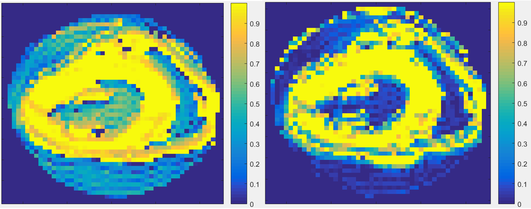
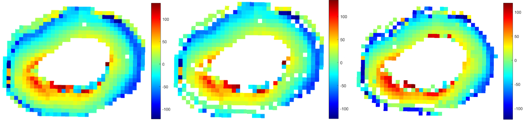
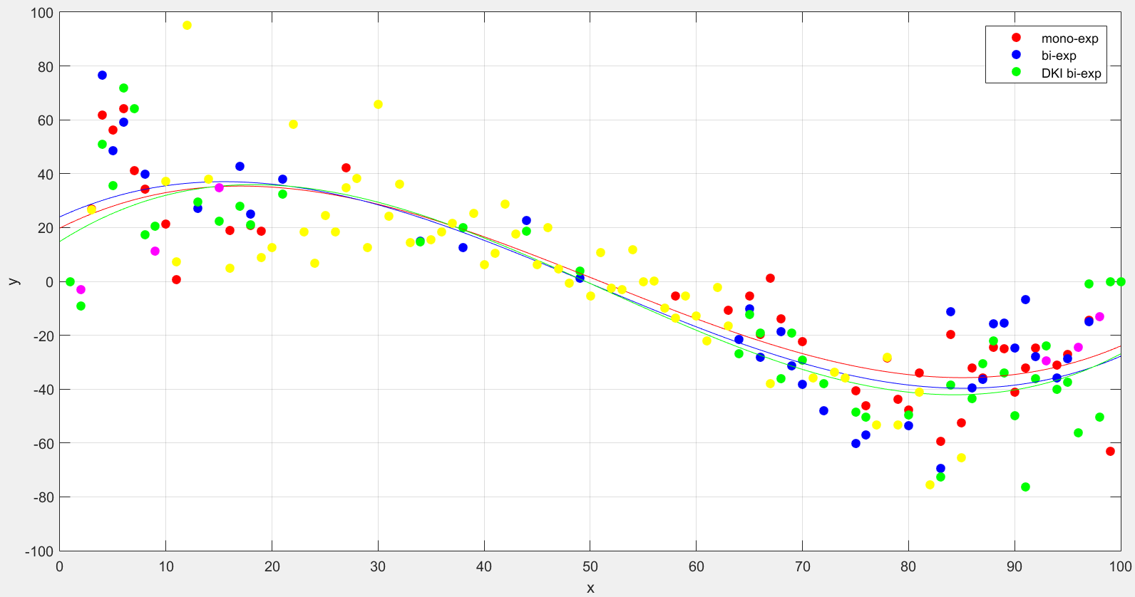
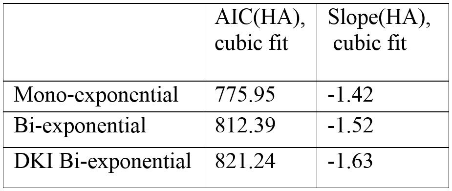
Figure1 shows the volume fraction calculated throughout the specimen for each of the bi-exponential diffusion models. Figure2 and 3 represent the HA calculated from each from the mono-, bi- and DKI bi- exponential models. The slope of a cubic fit to the HA across the myocardial wall was higher in the bi-exponential models and highest in the non-Gaussian model (Figure3 and 4). The Akaike information criterion (AIC) provided an indication of the quality of the fit.
Discussion
In Figure1, the DKI bi-exponential model provided a better estimate of the volume fraction. By accounting for the non-Gaussianity of the formalin diffusivity at high b-value, the DKI-bi model was able to more effectively isolate the formalin. A number of voxels around the edge of the container were identified as having a high percentage of tissue, resulting from the presence of susceptibility artefacts in those voxels. Accounting for the volume fraction led to improved delineation of tissue and formalin within the manually selected mask, especially at tissue boundaries (Figure2). Eliminating formalin-dominant voxels from the manually selected mask resulted in increased HA slope in bi-exponential models (Figure3 and 4). However, the curvature of the cubic fit privileges lower helix angle at the endo- and epicardium where partial volume effects are more likely. Thus, the AIC score performs better when using the mono-exponential model (Figure4). The HA fitting and its slope are dependent on the partial volume effects which can be accommodated by an appropriate diffusion model. In previous studies5, the maximum b-values used were limited to 1500s/mm2 as the models used did not account for the non-Gaussian effects at higher b-values. By including the higher b-values from the CUSP sampling9, and using a bi-exponential that accounts for non-Gaussian diffusion, the signals from fast diffusion components are better identified.Conclusion
The combination of high b-values and a non-Gaussian, bi-exponential diffusion model provides a method to better isolate the fast diffusion components. This method calculates tissue fraction in voxels affected by partial volume effects and can be used to more accurately define tissue masks, allowing automatic segmentation of the myocardium. In fixed heart specimens, this technique leads to better estimates of cardiac diffusion metrics, such as helix angle. For in vivo cardiac diffusion examinations, this technique can be adapted to account for the partial volume effects encountered between the myocardium and blood pool.Acknowledgements
This work was supported by the National Heart Foundation in New Zealand.References
1. Grebenkov DS: Use, Misuse, and Abuse of Apparent Diffusion Coefficients. Concepts Magn Reson Part A 2010; 36A:24–35.
2. Niendorf T, Dijkhuizen RM, Norris DG, van Lookeren Campagne M, Nicolay K: Biexponential diffusion attenuation in various states of brain tissue: implications for diffusion-weighted imaging. Magn Reson Med 1996; 36:847–857.
3. Mulkern R V., Haker SJ, Maier SE: On high b diffusion imaging in the human brain: ruminations and experimental insights. Magn Reson Imaging 2009; 27:1151–1162.
4. Pasternak O, Sochen N, Gur Y, Intrator N, Assaf Y: Free water elimination and mapping from diffusion MRI. Magn Reson Med 2009; 62:717–730.
5. Pasternak O, Shenton ME, Westin C-F: Estimation of Extracellular Volume from Regularized Multi-shell Diffusion MRI. Springer, Berlin, Heidelberg; 2012:305–312.
6. Jensen JH, Helpern JA, Ramani A, Lu H, Kaczynski K: Diffusional kurtosis imaging: The quantification of non-Gaussian water diffusion by means of magnetic resonance imaging. Magn Reson Med 2005; 53:1432–1440.
7. Jensen JH, Helpern JA: MRI quantification of non-Gaussian water diffusion by kurtosis analysis. NMR Biomed 2010; 23:698–710.
8. Veraart J, Poot DHJ, Van Hecke W, et al.: More accurate estimation of diffusion tensor parameters using diffusion kurtosis imaging. Magn Reson Med 2011; 65:138–145.
9. Scherrer B, Warfield SK: Parametric Representation of Multiple White Matter Fascicles from Cube and Sphere Diffusion MRI. PLoS One 2012; 7:e48232.
10. Cheng J, Shen D, Yap P-T, Basser PJ: Novel Single and Multiple Shell Uniform Sampling Schemes for Diffusion MRI Using Spherical Codes. Med Image Comput Comput Interv - MICCAI 2015 2015.
Figures