1589
Reproducibility of Diffusion Tensor Imaging Data between Morning and Evening Scans1Higher Institute of Bioscience of Paris (ISBS), Paris, France, 2MRC/UCT Medical Imaging Research Unit, Division of Biomedical Engineering, University of Cape Town, Cape Town, South Africa, 3Department of Human Biology, University of Cape Town, Cape Town, South Africa, 4Massachusetts General Hospital, Boston, MA, United States, 5Cape Universities Body Imaging Centre (CUBIC-UCT), Cape Town, South Africa
Synopsis
Diffusion Tensor Imaging (DTI) is widely used to study brain white matter integrity. However, instability of the MRI scanner including heating of the iron plates in the shim trays or physiological changes during the day may influence DTI indices. The aim of this work was to evaluate DTI parameters through scans performed at two different times of the day, early morning and late
Introduction
Diffusion encoding can be influenced by other factors not related to microstructural integrity that cause signal dephasing. For example, Alhamud et al. (2015) [1] previously reported frequency drifts in the scanner’s central frequency of roughly 30 Hz over 5 consecutive diffusion tensor imaging (DTI) acquisitions. Jiang et al. (2014) [2] in studies of diurnal microstructural alterations found significant changes in all DTI indices; however, their work did not include correction for B0 distortion and scans were only performed on the same day. In this work, we aimed to study over six days the influence of scan time on DTI parameters, comparing data acquired directly after the scanner is switched on (5:30 am; pre-heating status) to just before the scanner is switched off (6:00 pm; heated status), following a series of daily scans.Methods
DTI data were acquired from each of three healthy adults early in the morning (immediately after the scanner was switched on) and again in the late afternoon (after all daily scans were done) on six different days. A total of 18 morning scanning sessions, and 18 afternoon scanning sessions were performed. Scans were performed on a 3T Siemens Skyra with a 32 channel head coil. In each scanning session, two DTI acquisitions were performed with opposite phase encoding directions (i.e., anterior (A)–posterior (P) and PA) to correct for distortion in the B0 field using a twice-refocused SE-EPI sequence [3]: TR/TE 9800/92ms; 74 slices; 2x2x2 mm3; 30 diffusion directions with b=1000 s/mm2 and five non-weighted b=0 s/mm2 volumes.
DTI data preprocessing was performed using TORTOISE [4], including motion and eddy current correction, as well as EPI distortion corrections. The two DTI acquisitions were averaged and DTI parameters generated, including fractional anisotropy (FA), mean diffusivity (MD), axial diffusivity (AD) and radial diffusivity (RD). Unweighted B0 images were co-registered to their own T1-weighted structural image using linear and nonlinear coregistration algorithms in AFNI. All T1’s were co-registered to the MNI152 standard image with resolution 1.5x1.5x1.5 mm3. The DTI parameter maps were warped using the same transformation to achieve intra- and inter-subject alignment, and thresholded at an average FA>0.2 to ensure that only white matter was included.
Voxelwise group comparisons of DTI parameters were performed using FSL-randomise [5] to identify clusters showing significant differences (morning versus afternoon scans). We only report clusters that survived cluster size correction at p<0.05 [6].
Results
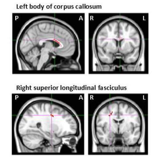
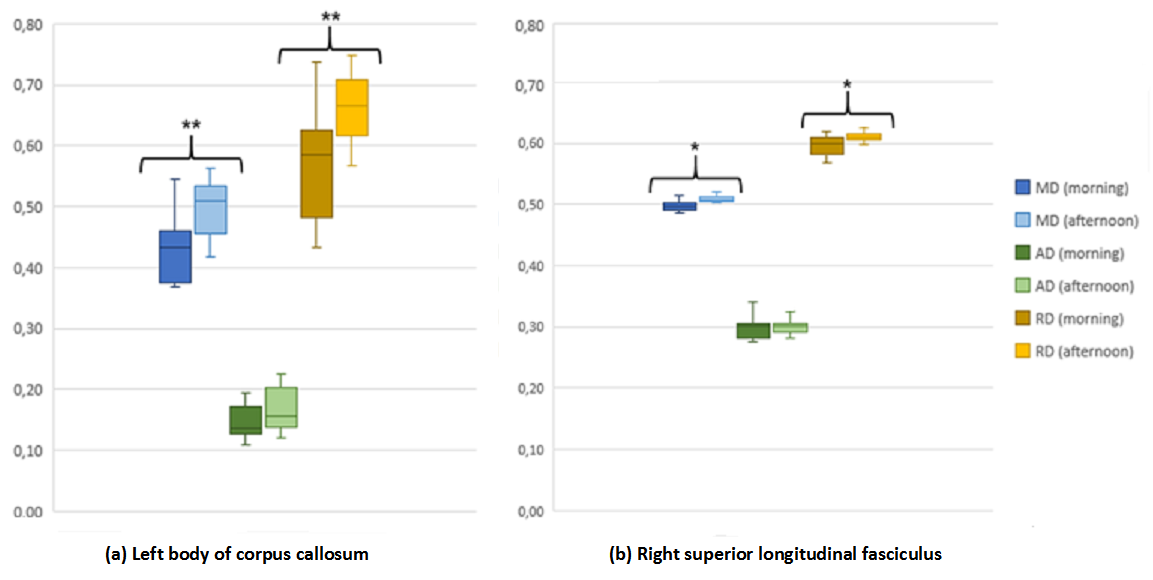
FA maps showed no significant differences between morning and afternoon scans. Higher MD was seen in the morning scans compared to the afternoon scans in two regions, namely left body of corpus callosum and right superior longitudinal fasciculus (Figure 1). Comparison of the cluster means of MD, AD, and RD for morning and afternoon scans in each of these regions (Figure 2a,b) revealed that MD differences were attributable to differences in RD.Discussion
Our results suggest that performing MRI scans at different times of day may affect MD and RD measures regionally, while FA and AD measures appear to be more robust to timing of scans. Our findings support those of Jiang et al. (2014) [2] who reported that ADC and RD were significantly increased in the morning. In contrast, they also found a similar trend in FA which was not observed in this work. Observed diffusivity alterations may also, in part, be related to heating of shim irons and this requires further investigation.Conclusion
For longitudinal study or studies that require repeated scans, it may be necessary to perform scans at similar times of day to avoid any variability in DTI indices that may arise from physiological changes. Our findings suggest that FA and AD may offer better reproducibility than MD and RD.Acknowledgements
The National Research Foundation of South Africa (NRF), Thuthuka grant TTK150612119380.References
1. Alhamud A, Taylor PA, van der Kouwe AJ, Meintjes EM. Real-time measurement and correction of both B0 changes and subject motion in diffusion tensor imaging using a double volumetric navigated (DvNav) sequence. Neuroimage. 2016 Feb 1;126:60-71.
2. Jiang C, Zhang L, Zou C, Long X, Liu X, Zheng H, Liao W, Diao Y. Diurnal microstructural variations in healthy adult brain revealed by diffusion tensor imaging. PLoS One. 2014 Jan 6;9(1):e84822.
3. Reese TG, Heid O, Weisskoff RM, Wedeen VJ. Reduction of eddy-current-induced distortion in diffusion MRI using a twice-refocused spin echo. Magn Reson Med. 2003;49(1):177–182.
4. Pierpaoli C, Walker L., Irfanoglu MO, Barnett A, Basser P, Chang L-C, Koay C, Pajevic S, Rohde , Sarlls J, Wu M. TORTOISE: an integrated software package for processing of diffusion MRI data, ISMRM 18th annual meeting, 2010, Stockholm, Sweden, #1597.
5. Winkler AM, Ridgway GR, Webster MA, Smith SM, Nichols TE. Permutation inference for the general linear model. NeuroImage, 2014, 92, 381-397.
6. Forman SD, Cohen JD, Fitzgerald M, Eddy WF, Mintun MA, Noll DC. Improved assessment of significant activation in functional magnetic resonance imaging (fMRI): use of a cluster-size threshold. Magnetic Resonance Medicine, 1995, 33, 636–647.
Figures