1566
Predictive Value of Two-tensor Unscented Kalman Filter Tractography in the Reconstruction of the Arcuate Fasciculus (AF) in Patients with Gliomas Involving Eloquent Language Areas1Department of MRI, the First Affiliated Hospital of Zhengzhou University, Zhengzhou, China, 2Siemens Healthcare, Scientific marketing, Beijing, China, 3Department of Neurosurgery, the First Affiliated Hospital of Zhengzhou University, Zhengzhou, China
Synopsis
This study aimed to preliminarily investigate the postoperative changes of AF in glioma patients detected by two-tensor UKF tractography from the perspective of the usefulness as a reference for postoperative recovery of language functions. The postoperative changes of AF were evaluated chronologically in relation to postoperative changes in language functions after surgery. Our study preliminarily shows that postoperative changes in the long segment of the left AF detected by two-tensor UKF tractography may be a predicting factor for postoperative language functional outcomes. Postoperative changes in the long and posterior segment of the left AF may be related with the language comprehending and repeating ability in glioma patients.
Introduction
Disturbance of arcuate fasciculus (AF) in the dominant hemisphere is thought to be associated with language-processing disorders[1, 2]. Although the AF can be visualized in vivo with DTI tractography, its involvement in language related functional processes has been rarely shown in longitudinal studies for brain surgery. In addition, due to the drawbacks of the DTI technique, such as fiber-crossing effects, partial-volume effects and abnormal diffusivity of white matter in the intra- and peri-tumoral areas, the tractography result based on the conventional single-tensor model is inaccurate[3, 4]. To address these problems, we applied a novel tractography technique based on a two-tensor unscented Kalman filter (UKF) algorithm, named as two-tensor UKF tractography[5]. In this study, we employed this technique in a longitudinal research for patients with gliomas involving eloquent language areas before and after surgery. Specifically, we quantified the restorative condition of AF by comparing the pre- and postoperative AF volume, and correlated the AF volume with the language function, in order to provide some references for the recovery of language function of giloma patients from the perspective of language-related white matter change.Method
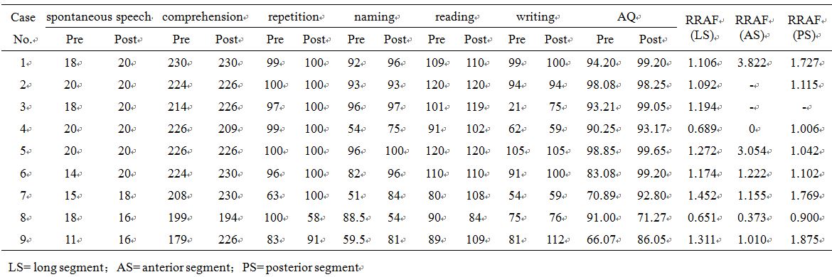
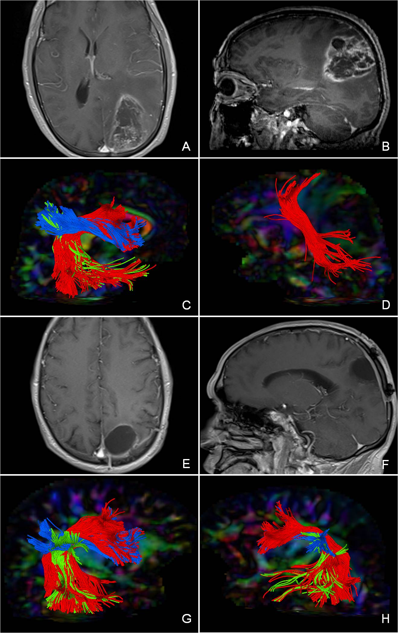
Nine right-handed patients with gliomas involving eloquent language areas successfully postoperative follow-up were enrolled, and all of them received 3D MPRAGE, T2-FLAIR and DTI scanning on a 3T intraoperative magnetic resonance imaging (iMRI) scanner (MAGNETOM Verio, Siemens Healthcare, Erlangen, Germany). 3D MPRAGE images were acquired with the following parameters: TR=1900 ms, TE=2.93 ms, flip angle=9°, matrix size=256×215, slice thickness=1 mm. T2-FLAIR images were acquired with the following parameters: TR=9000 ms, TE=96 ms, TI=2500 ms, flip angle=150°, matrix size=256×160, slice thickness=2 mm. DTI were acquired using SS-EPI sequence with the following parameters: 30 gradient directions evenly distributed on the sphere, b-value of 1000s/mm2, TR=9900 ms, TE=90 ms, NEX=2, matrix size=128×128, voxel size=2.0×2.0×2.0 mm3, slice thickness=2 mm. Two-tensor UKF tractography was calculated using the 3D slicer software and was applied to reconstruct the direct long segment, anterior fronto-parietal segment and posterior temporal-parietal segment of the bilateral AF. To account for the volume difference between pre- and postoperative conditions of the long, anterior and posterior segment of left AF, the relative ratio of AF (RRAF) was calculated in each patient according to the following formula: RRAF=(volume of the postoperative left AF/volume of the postoperative right AF)/(volume of the preoperative left AF/volume of the preoperative right AF). An RRAF greater than 1.0 indicated increased visualization of the postoperative left AF. Language assessment was conducted accompany with MRI scanning using the aphasia battery of Chinese, including spontaneous speech, comprehension, repetition, naming, reading, and writing. Aphasia quotient (AQ) was calculated from the score of the first four language functions and adopted to evaluate the severity of aphasia. To account for the different pre- and postoperative conditions, changes in language functions was calculated in each patient according to the following formula: substraction score in language function=(postoperative score in language function)-(preoperative score in language function).Result
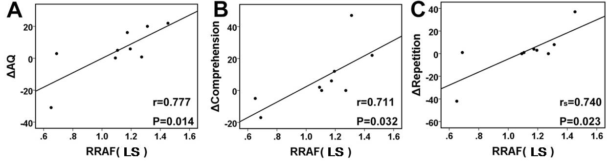
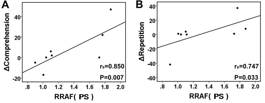
Postoperative RRAF of long segment of the left AF was positively correlated with substraction score in AQ (r=0.777, P=0.014), substraction score in comprehension (r=0.711, P=0.032) and substraction score in repetition (rs=0.74, P=0.023); Postoperative RRAF of posterior segment of the left AF was positively correlated with substraction score in comprehension (rs=0.850, P=0.007) and substraction score in repetition (rs=0.747, P=0.033).Discussion and Conclusion
The two-tensor UKF algorithm was designed to perform tractography within a Kalman filter framework using a mixture of two Gaussian tensors to model the signal. The local fiber orientations are traced using the estimation at previous positions to guide estimation at the current position. In a loop, the Kalman filter estimates the model at the current position, moves a step in the most consistnet direction, and then begins estimation again. Recursive estimation in this manner greatly improves the accuracy of resolving individual orientations and yield inherently smooth tracts despite the presence of noise and uncertainty. This tractography algorithm allows restruction of tracts that pass through branching and crossing fiber regions of the human brain. Our study shows that two-tensor UKF tractography can relatively completely visualize and quantitate AF in patients with gliomas involving eloquent language areas, thereby displaying the restorative condition of the postoperative AF. This study preliminarily shows that increasing the volume of the long segment of the left AF may be a predictor of a well restoration of postoperative language function. The restoration of postoperative long and posterior segment of the left AF may be helpful for improving language comprehending and repeating ability of patients. However, due to the small number of cases in this study, it is necessary to increase the sample size to further confirm the reliability of predictive value of AF restoration in the reversion of language function.Acknowledgements
No acknowledgement found.References
[1] Catani M, Mesulam M. The arcuate fasciculus and the disconnection theme in language and aphasia: history and current state[J]. Cortex, 2008, 44(8): 953-61.
[2] Bernal B, Ardila A. The role of the arcuate fasciculus in conduction aphasia[J]. Brain, 2009, 132(Pt 9): 2309-16.
[3] Zhang H, Wang Y, Lu T, et al. Differences between generalized q-sampling imaging and diffusion tensor imaging in the preoperative visualization of the nerve fiber tracts within peritumoral edema in brain[J]. Neurosurgery, 2013, 73(6): 1044-53; discussion 1053.
[4] Kuhnt D, Bauer MH, Egger J, et al. Fiber tractography based on diffusion tensor imaging compared with high-angular-resolution diffusion imaging with compressed sensing: initial experience[J]. Neurosurgery, 2013, 72 Suppl 1: 165-75.
[5] Malcolm JG, Shenton ME, Rathi Y. Filtered multitensor tractography[J]. IEEE Trans Med Imaging, 2010, 29(9): 1664-75.
Figures