1560
Free Water Elimination Improves Tractography Through Multiple Sclerosis Lesions1Save Sight Institute, Sydney Medical School, University of Sydney, Sydney, Australia, 2Faculty of Medicine and Health Sciences, Macquarie University, Sydney, Australia, 3Brain and Mind Centre, University of Sydney, Sydney, Australia, 4Sydney Neuroimaging Analysis Centre, Sydney, Australia, 5Brigham and Women’s Hospital, Harvard Medical School, Boston, MA, United States
Synopsis
Axonal loss within chronic MS lesions is typically accompanied by increase of extra-cellular space. Reduction of anisotropy caused by this excessive extra-cellular water may limit the ability of tractography techniques to accurately detect fibre bundles. The aim of this study was to examine if application of free water elimination (FWE) algorithm may improve deterministic tractography through MS lesions. We show that elimination of free water markedly increases detection of lesional fibre bundles. While this effect was observed in the majority of lesions, it was more apparent in lesions with small initial number of fibres and in lesions categorised as severely damaged.
Introduction
DTI-based tractography is a sensitive clinical research tool used to understand connectivity and microstructural brain changes in neurodegenerative disorders.(1) Probabilistic tractography algorithms typically generate large number of fibres, but require pre-existing knowledge of fibre-tract anatomy and manual cleaning of the ‘erroneous’ fibres. Deterministic tractography is less susceptible to contamination, but in pathologically-altered areas of white matter is more affected by decreased anisotropy. In MS lesions, the combination of post-inflammatory gliosis, demyelination and axonal loss causes significant increase of extra-cellular water and, as a consequence, reductions of anisotropy. This often leads to partial failure of tractography algorithms. However, as extracellular water content within a lesion is thought to be isotropic, confounding extracellular water signals in a lesion can potentially be removed. Algorithms for removing excessive extracellular free water from brain tissue have been successfully applied to other neurodegenerative conditions.(2-5) In this current study we investigate if Free Water Elimination (FWE) can improve deterministic tractography through MS lesions and assess how Free Water Elimination is affected by the degree of lesional damage.
Method
The following sequences were acquired using a 3T GE Discovery MR750 scanner (GE Medical Systems, Milwaukee, WI) as described in detail previously: (6)
- Pre- and post- contrast (gadolinium) Sagittal 3D T1
- FLAIR CUBE;
- Whole brain 64-directions diffusion weighted imaging with 2mm isotropic acquisition matrix.
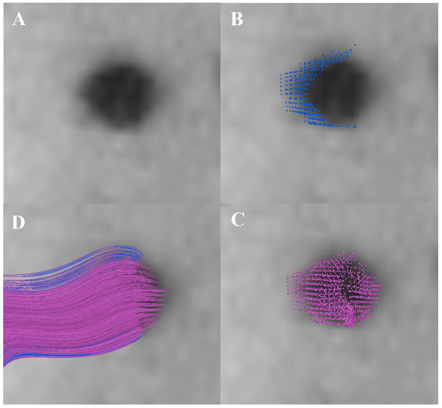
141 randomly chosen lesions with varying degrees of T1 hypointensity were selected from 36 RRMS patients. Deterministic tractography was performed before and after application of FWE algorithm using lesion mask as ROI 1. Individual lesion masks were generated using ITK-SNAP, including 53 ‘moderate’ lesions from 25 subjects (mean T1 hypointensity ≥ 50% grey matter) and 88 ‘severe’ lesions from 28 subjects (defined as mean T1 hypointensity < 50% grey matter). MrDiffusion’s deterministic tractography (Stanford University) was used to generate fibres from ROIs with the following parameters (step size: 1mm, angle threshold: 30, FA threshold: 0.15, length threshold: 20mm). The generated fibres were visualised with Quench.
Results and Discussion
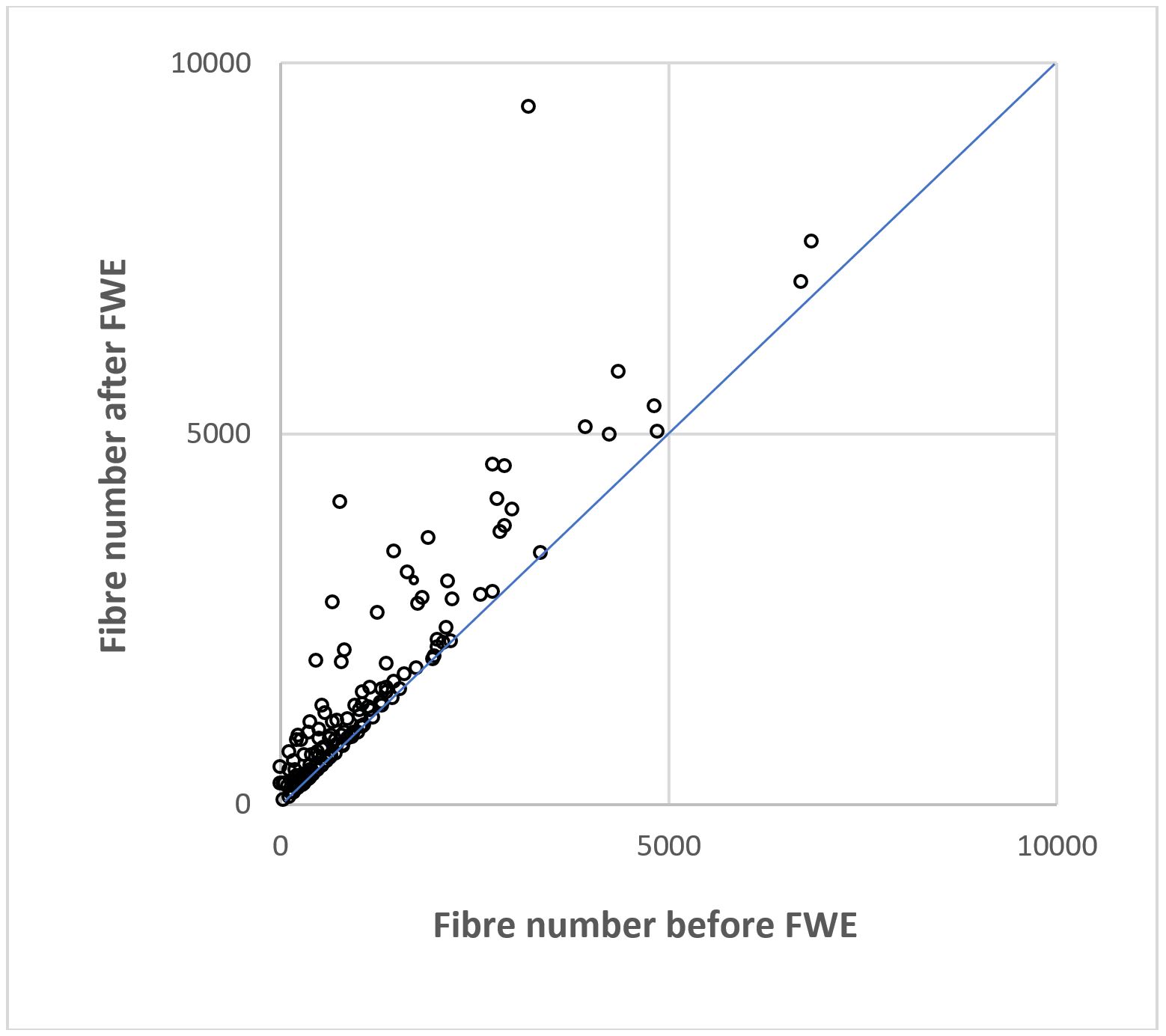
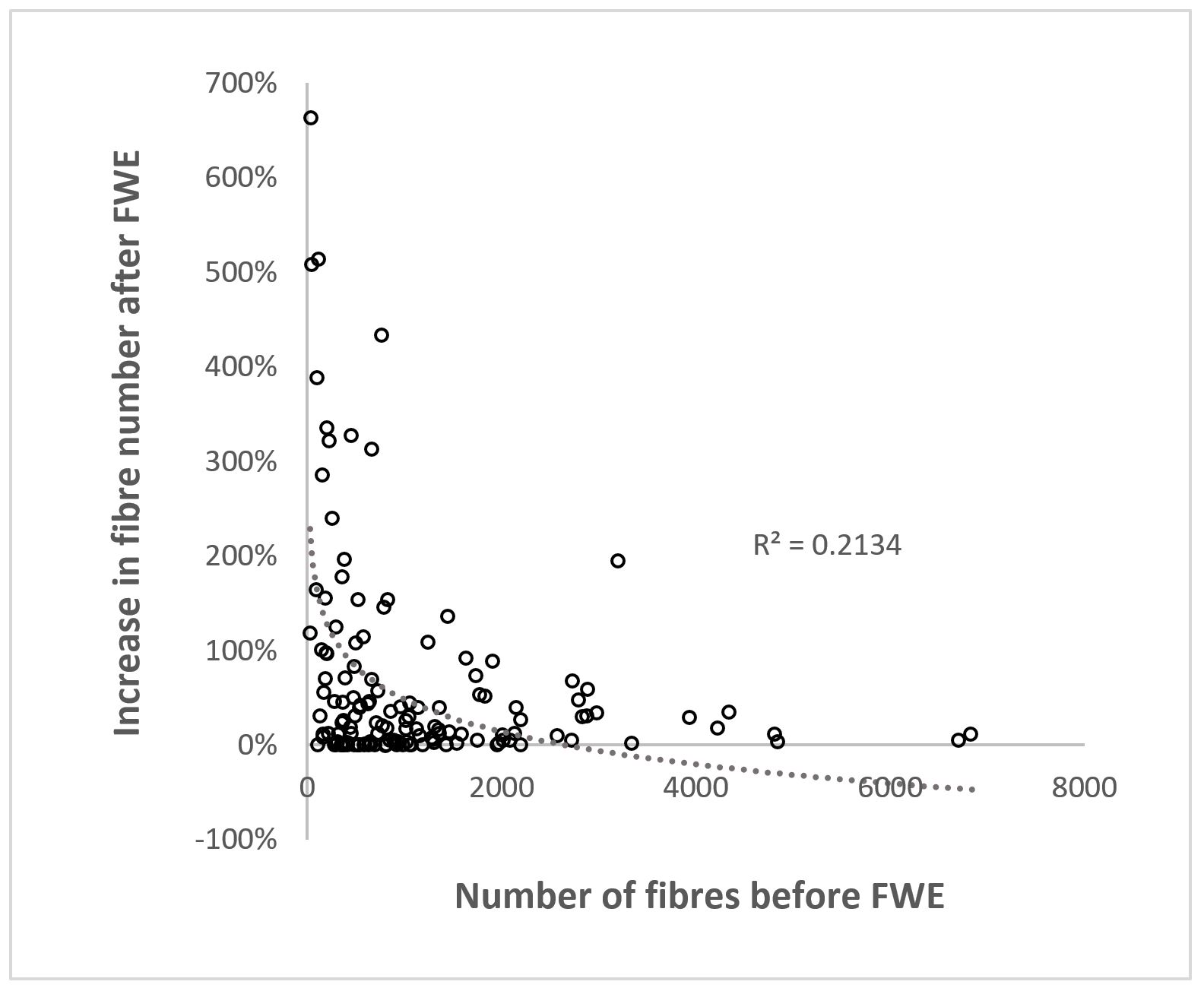
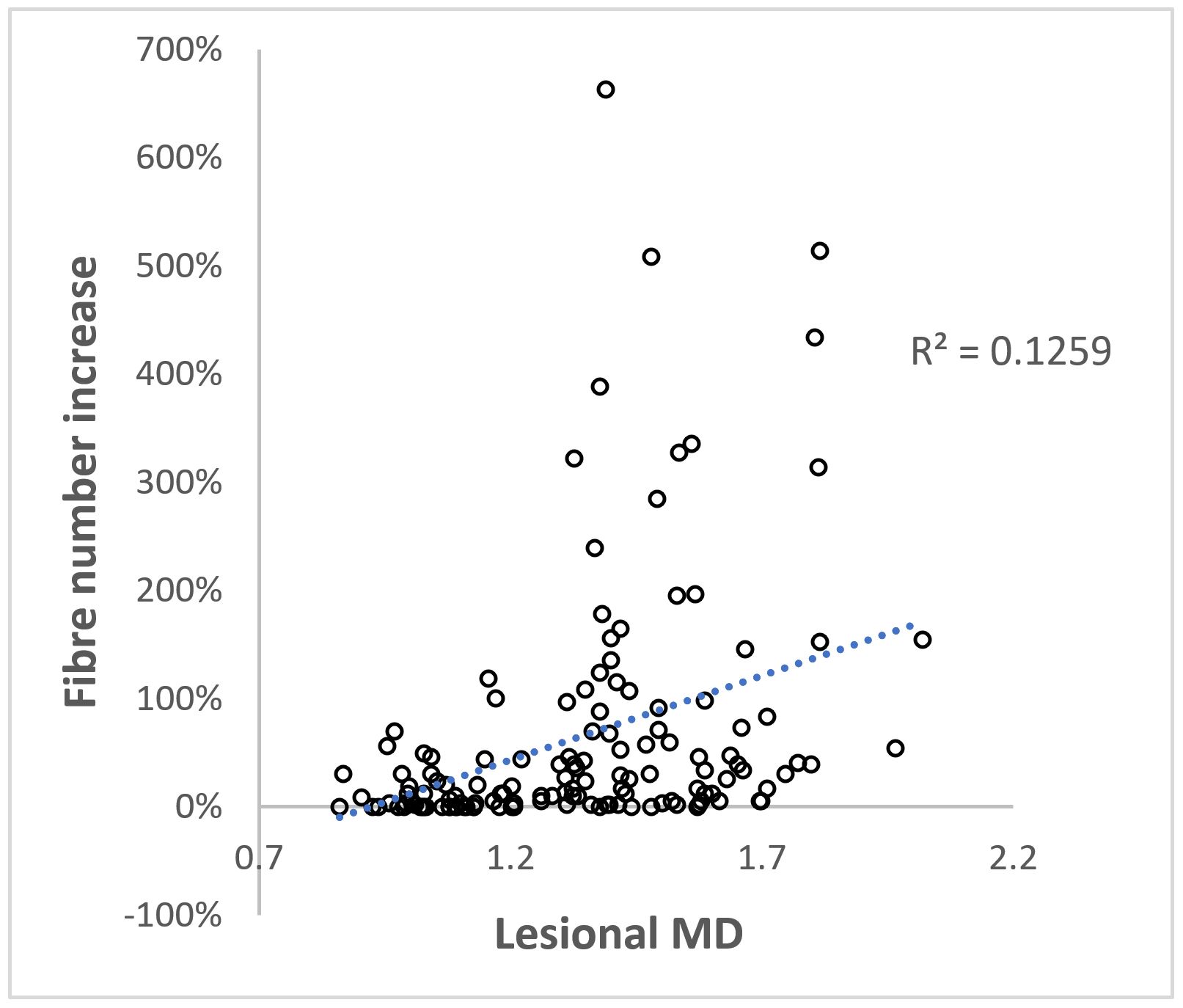
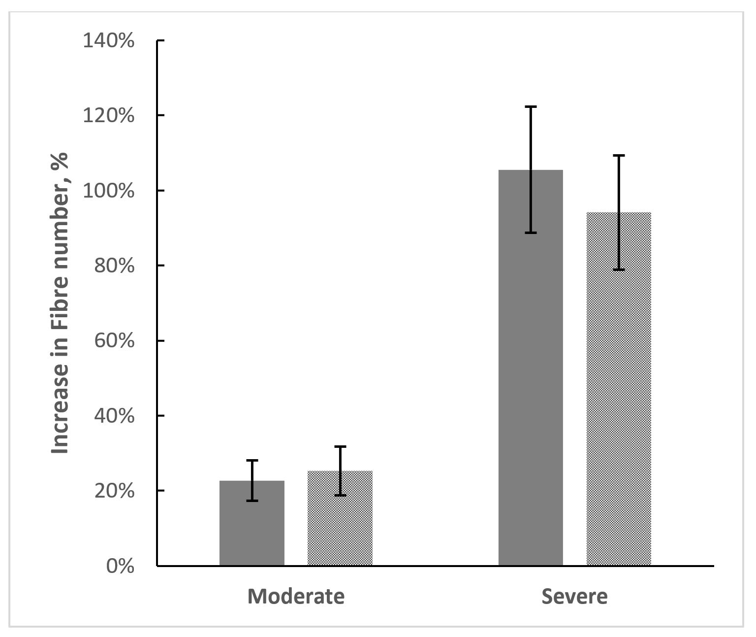
There was an overall significant increase in the number of lesional fibres defined as fibres traversing through the lesion after application of the FWE algorithm (p = 1.3e-10 using a two-tailed paired t-test). (Fig.1) Increase was seen in the majority of cases (114/141). (Fig. 2) The relative increase in fibre count observed after FWE was more apparent in lesions with small initial number of fibres. (Fig. 3) The improvement was also more marked in severely damaged lesions (as measured by T1 hypointensity or lesional Mean Diffusivity). Thus, there was significant correlation between T1 hypointensity (or lesional MD) and relative increase in fibre numbers (R=0.3 and 0.46 respectively). (Fig. 4) When lesions were grouped into severely or moderately destructive based on T1-hypointensity or MD, there was significant difference between groups in relation to tractography improvement (106 % vs 23% and 94% vs 25%). (Fig. 5) There were two cases where without FWE no fibres from a lesion could have been generated. Upon FWE, 521 and 291 fibres were identified, respectively.Conclusion
The FWE process significantly improves deterministic tractography through MS lesions, particularly in the cases of severe tissue damage and, therefore, should become an essential component in fibre-based diffusivity analysisAcknowledgements
No acknowledgement found.References
- Assaf Y, Pasternak O. Diffusion tensor imaging (DTI)-based white matter mapping in brain research: A review. J Mol. Neurosci. 2008;34:51-61
- Pasternak O, Sochen N, Gur Y, et al. Free water elimination and mapping from diffusion MRI. Magn. Reson. Med. 2009;62:717-730.
- Mandl R, Pasternak O, Weipke C, et al. Comparing free water imaging and magnetization transfer measurements in schizophrenia. Schizophr Res. 2015;161(1):126-132.
- Maier-Hein K, Westin C, Shenton M, et al. Widespread white matter degeneration preceding the onset of dementia. Alzheimers Dement. 2015;11(5):485-493.
- Bergamino M, Pasternak O, Farmer M, et al. Applying a free-water correction to diffusion imaging data uncovers stress-related neural pathology in depression. Neuroimage Clin. 2016;10:336-342.
- Klistorner A, Wang C, Fofanova V, et al. Diffusivity in multiple sclerosis lesions : At the cutting edge? NeuroImage Clin. 2016;12:219-226.
Figures