1555
Bundle-specific tractography using voxel-wise orientation priors1Computer Science, Université de Sherbrooke, Lac-Etchemin, QC, Canada, 2Computer Science, Université de Sherbrooke, Sherbrooke, QC, Canada, 3Groupe d'Imagerie Neurofonctionnelles, Institut des Maladies Neurodégénératives (GIN-IMN) - UMR 5293, CNRS, CEA, Université de Bordeaux, Bordeaux, France
Synopsis
Diffusion tractography allows the investigation of white matter (WM) pathways of interest. However, to cover the full spatial extent of the desired bundles, tractography requires a large amount of streamlines (millions) to be generated. In this work, we developed a bundle-specific tractography algorithm using voxel-wise orientation priors. Our method aims to be more efficient than a classical whole brain tractography and increase the quality of virtual WM dissection.
Purpose
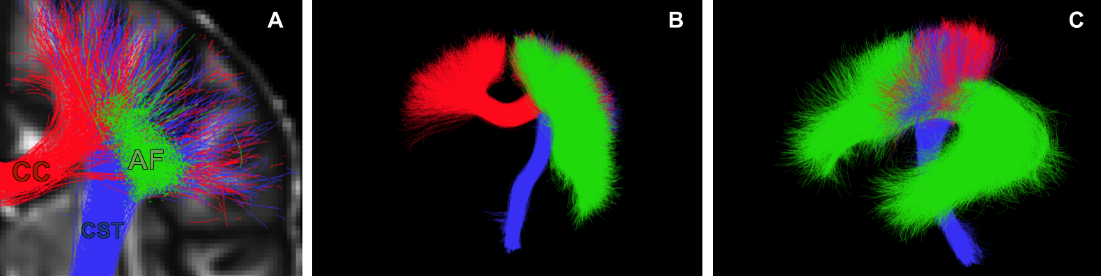
Reconstruction of white matter (WM) bundles using tractography is a difficult task1,2. It often requires the generation of a large number of streamlines in order to cover the spatial extent of a desired bundles3. In this work, we developed a bundle-specific tractography algorithm to avoid a full brain tractography. We propose a novel local orientation enhancement methodology, inspired by TOD, based on white matter fascicle priors. This method can overcome reconstruction difficulties in curved or crossing regions4, such as the one illustrated in Figure 1. These enhancements yield better spatial coverage and increases the quality of the fanning which helps to accurately represent the fascicle. As such, this bundle-specific approach is an alternative to the classical whole brain tractography for researchers examining certain bundles of interest (BOI).
Methods
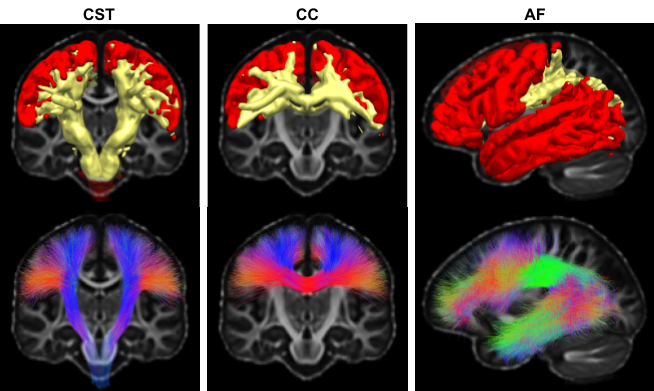
Our proposed approach is composed of 3 steps. First, a template that represents the general shape and position of the BOI needs to be created from existing bundle. The streamlines need to be free of loop or outlier and their endpoints need to be representative of the termination of the BOI. Then, the template is used on a new subject, by bringing the template into the subject native space. Using the information from the template, seeding and tracking masks can be created (as seen in Figure 2) as well as orientation priors. Our approach uses orientation priors to modify the weighting according to the general apriori direction in the voxel.
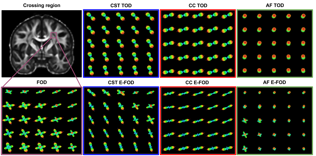
To create our orientation priors we compute the voxel-wise local orientation histogram from nearby segments of streamlines5. In our work, directions observed in the BOI template are used to enhance the fiber orientation distribution (FOD) to track specifically the same BOI. To create an orientational insight without imposing a strong orientation prior, the template TOD is constructed using collection from delta function in spherical harmonic of order two.Then, at each voxel, FOD can be re-weighted by computing a point-wise multiplication of the FOD sphere with the orientation priors followed by a normalization. The consequence is that the most well-aligned lobe with our orientation priors will have its values increase in proportion to the others, resulting in an increased probability of following that general direction. This operation does not create an artificial lobe if it is not existent on the original FOD.
In this work, probabilistic particles filtering tracking6 was used to reconstruct 5 BOI, the Arcuate Fasciculus (AF), the Corticospinal Tract (CST) and a sub-portion of the Corpus Callosum (CC).
Results
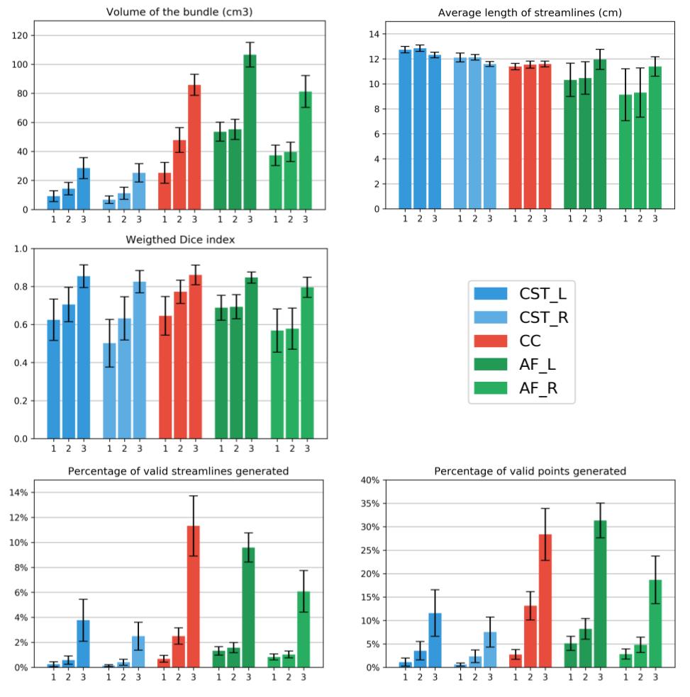
Evaluation was performed using the following measures: bundle volume, weighted-Dice coefficient6, average streamlines length, percentage of valid streamlines and computational performance. The percentage of valid streamlines is the proportion of generated streamlines respecting the anatomical definition of a bundle from our expert.
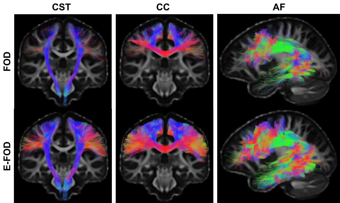
As illustrated in Figure 4, reconstructed BOI from the standard tracking shows some degree of fanning, but the number of streamlines generated was not sufficient to fully recover the spatial extent of bundles. All BOI reconstructed using the masking priors and the enhanced-FOD show an increase in both bundle coverage and quality of the fanning. For the CST, the lateral projection of the pre/postcentral gyri is more represented. The same thing can be observed for the CC. As for the AF the spatial extent in the frontal and temporal lobe is increased, but streamlines are also reaching laterally into the gyri. The quantitative impact of the improved fanning directly impact the volume of the reconstructed bundle.
Most tractograms contain millions of streamlines to guarantee a stable volume as well as a stable overlap among subjects3. Weighted-Dice index of each bundle also increases when using our method. Our proposed method not only increases the volume, but we also obtain a more coherent overlap between our subjects. Figure 5 shows all measures for each of the 5 bundles, the 1/2/3 bars represent 3 different tracking paramaters :
- Bundle-specific seeding, WM mask, original FOD
- Bundle-specific seeding, bundle-specific mask, original FOD
- Bundle-specific seeding, bundle-specific mask, bundle-specific FOD
Conclusion
We have developed a template-based FOD enhancing method to overcome reconstruction difficulties. Each step of the map creation is straightforward, but their creation using a WM atlas composed of streamlines is a novel approach. This automated approach uses information extracted from streamlines of a template to enhance the local modeling in the desired direction using the apriori orientation distribution. Our approach greatly accelerates tractography reconstruction of specific bundles and facilitates future manual dissections7. We have shown that our approach improves the spatial coverage and increases the quality of the fanning in crossing regions, while reducing computational need. Our proposed method can help neuroanatomists to explore the structural connectivity of each subjects with more confidence.
Acknowledgements
We would like to thank Jasmeen Sidhu and Michael Paquette from the Sherbrooke Connectivity Imaging Lab (SCIL) for their help during the early phase.The authors would like to thank the Fonds de recherche du Québec – Nature et technologies (FRQNT) and NSERC Collaborative Research and Training Experience Program in Medical Image Analysis (CREATE-MIA) for funding this research. We also thank the Université de Sherbrooke institutional chair in neuroinformatics for their support.References
Jbabdi, S., Johansen-Berg, H., 2011. Tractography: where do we go from here? Brain connectivity 1, 169–183.
Chamberland, M., Scherrer, B., Prabhu, S.P., Madsen, J., Fortin, D., Whittingstall, K., Descoteaux, M., Warfield, S.K., 2017. Active delineation of meyer’s loop using oriented priors through magnetic tractography (magnet). Human Brain Mapping 38, 509–527.
Tournier, J.D., Calamante, F., Connelly, A., 2012. MRtrix: Diffusion tractography in crossing fiberregions. International Journal of Imaging Systems and Technology 22, 53–66.
Gauvin, A., Petit, L., Descoteaux, M., 2016. Data: Achieving volume saturation of streamline bundles in tractography, in: Proceedings of the International Society of Magnetic Resonance in Medicine, p. 2500.
Dhollander, T., Emsell, L., Hecke, W.V., Maes, F., Sunaert, S., Suetens, P., 2014. Track orientation density imaging (todi) and track orientation distribution (tod) based tractography. NeuroImage 94, 312 – 336.
Girard, G., Descoteaux, M., 2014. Towards quantitative connectivity analysis: reducing tractography biases. NeuroImage 98, 266–278.
Cousineau, M., Jodoin, P.M., Garyfallidis, E., Ct, M.A., Morency, F.C., Rozanski, V., GrandMaison, M., Bedell, B.J., Descoteaux, M., 2017. A test-retest study on parkinson’s ppmi dataset yields statistically significant white matter fascicles. NeuroImage: Clinical 16, 222 – 233.
Catani, M., De Schotten, M.T., 2008. A diffusion tensor imaging tractography atlas for virtual in vivodissections. Cortex 44, 1105–1132.
Figures