1540
MP2RAGE-Compressed Sensing for fast metastasis detection and characterization in mice1CRMSB UMR5536, CNRS-Univ.Bordeaux, Bordeaux, France, 2CRMBM UMR7339, CNRS/Aix-Marseille Univ., Marseille, France
Synopsis
In order to detect and characterize metastases in preclinical studies, 3D T1 maps can be obtained with the MP2RAGE sequence. As high spatial resolution is required, the acquisition duration becomes prohibitive for the monitoring of metastases. Thus, acceleration via Compressed Sensing technique was achieved, necessitating a new undersampling scheme. T1 maps of the mouse whole brain were obtained in <1min. The T1 of brain metastases was not affected by CS acceleration. Then, ultra-high spatially resolved maps (130x125x141μm) were acquired without lengthening scan time, to detect early-growing metastases and accurately measure their volumes.
INTRODUCTION
Metastases are a major cause of death for cancer patients. T1 mapping using the MP2RAGE sequence can be employed to detect early stage metastases and characterize them longitudinally 1. However, the acquisition duration for 3D T1 mapping sequence remains prohibitive2 for routine use during the longitudinal monitoring of metastases. Among the techniques used to accelerate acquisition, the « Compressed Sensing » (CS) technique is of great interest, especially for preclinical studies, where the amount of receiving coils is considerably limited and high SNR is crucial. Consequently, we proposed a novel MP2RAGE sequence with a dedicated k-space sampling scheme for CS image reconstruction technique in order to obtain 3D T1 maps of the mouse brain at 7T. The benefit from the acceleration was demonstrated both to reduce acquisition duration and to increase the spatial resolution without lengthening scan time.MATERIALS AND METHODS
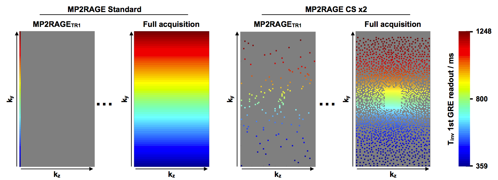
The encoding scheme of the standard Cartesian MP2RAGE sequence has been modified to include a variable density Poisson (VDPoisson) disk under-sampling distribution along the ky-kz axis. Briefly, instead of the standard MP2RAGE sequence, where each echo train is acquired with the same kz value during an MP2RAGETR, in the proposed implementation, the CS-MP2RAGE echo trains are spread in the ky-kz plan as shown in Figure 1. Consequently, one of the effects of the VDPoisson disk under-sampling is the accumulation of a broader range of effective Tinv at the centre of the ky axis independently of the acceleration factor (CS).
To evaluate the accuracy of the T1 measurements using the CS-MP2RAGE sequences, tubes containing increasing Gd-DTPA concentrations were used to generate T1 values from 792ms to 1979ms.
Five nu/nu mice were injected into the left ventricle of the beating heart with 100 μL suspension of 150 000 breast cancer cells (MDA-MB-231Br cells). They were scanned at different time points on a 7T Bruker BioSpec system (Ettlingen, Germany).
The MP2RAGE parameters were: Ti1/Ti2/MP2RAGETR= 800ms/2200ms/6250ms; FOV= 25x20x18; flip angle= 7°. Two spatial resolutions were acquired: Low (noted LR thereafter) with matrix=128x128x64 and High (noted HR thereafter) with matrix=192x160x128. Consequently, seven sequences were acquired:
LR : TE/TR = 3.253/7ms; BW = 29761Hz; 6min52s
LR CS2 : TE/TR = 3.253/7ms; BW = 29761Hz; 3min25s
LR CS4 : TE/TR = 3.253/7ms; BW = 29761Hz; 1min42s
LR CS6 : TE/TR = 3.253/7ms; BW = 29761Hz; 51s
HR : TE/TR = 3.072/8.056ms; BW = 50000HZ; 16 min 42 s
HR + CS2 : TE/TR = 3.072/8.056ms; BW = 50000Hz; 8 min 07 s
HR + CS4 : TE/TR = 3.072/8.056ms; BW = 50000Hz; 4 min 16 s
The 3D T1 maps were reconstructed using the Matlab software (MathWorks, USA). The Compressed-Sensing encoding scheme was used to performed a l1-ESPIRIT reconstruction with l1 wavelet regularization and total variation. This ESPIRIT-based parallel imaging and Compressed Sensing reconstruction was performed using the Berkeley Advanced Reconstruction Toolbox3.
RESULTS
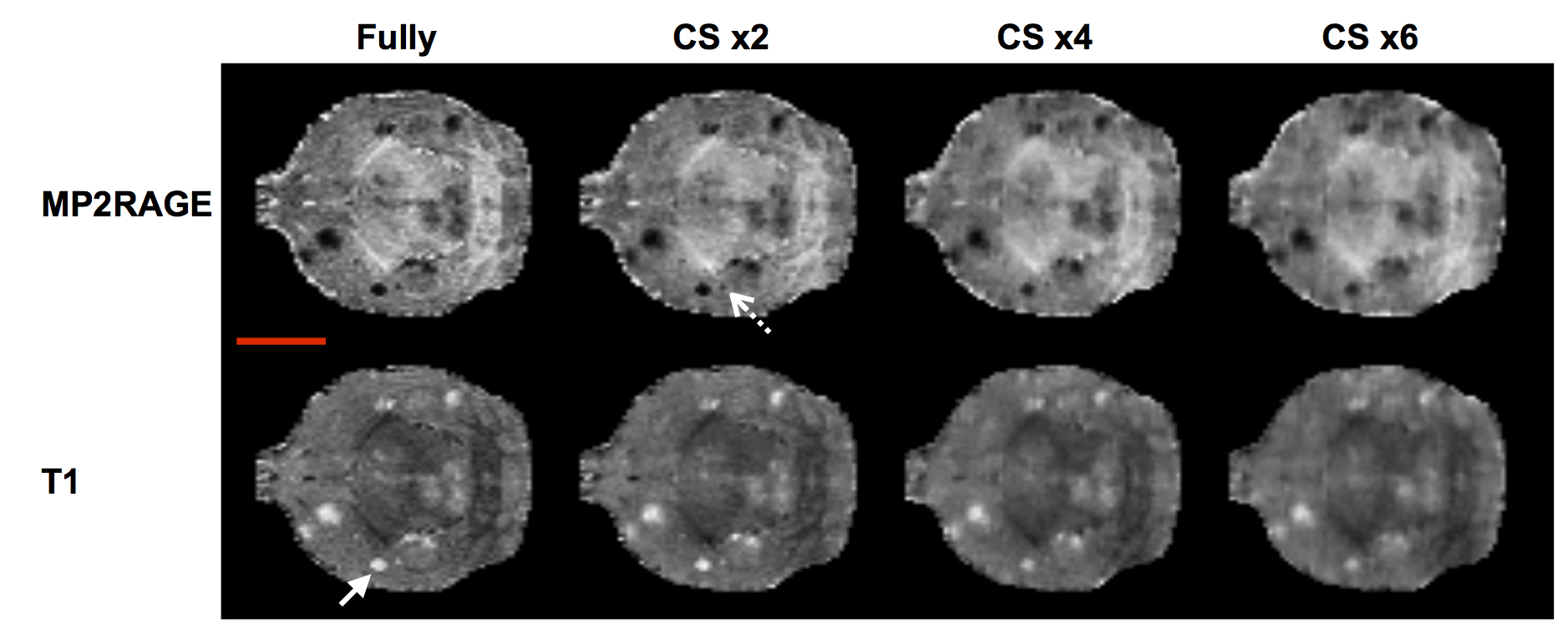
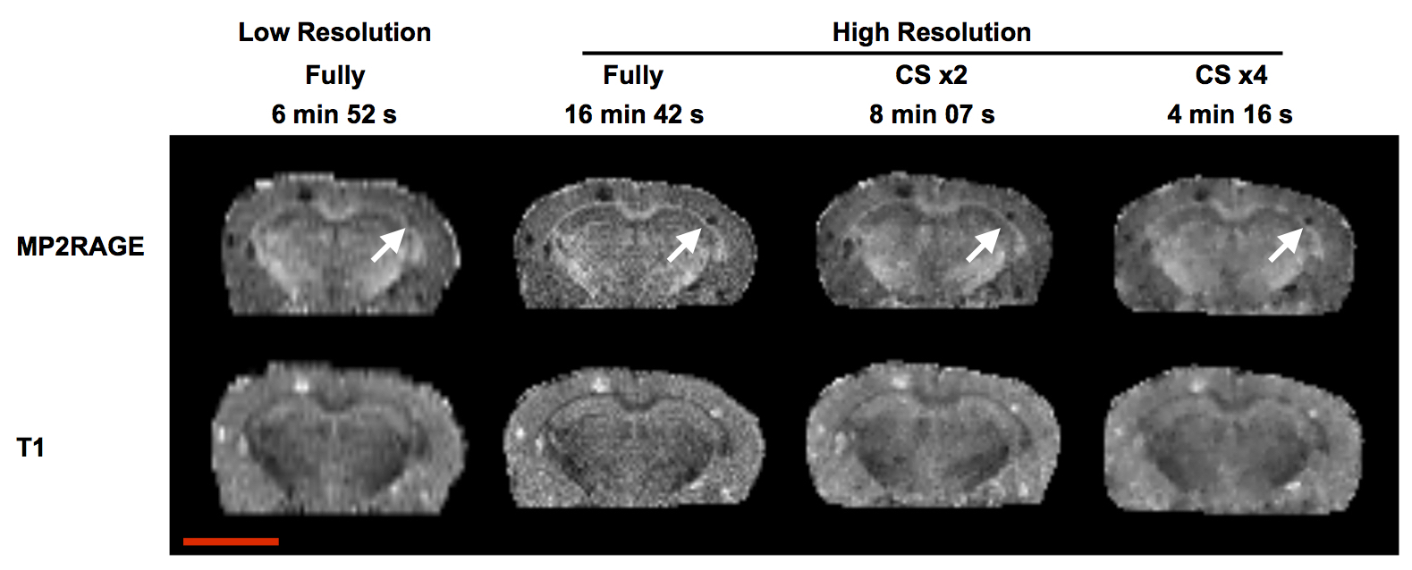
The T1 values of the different tubes obtained from the MP2RAGE-LR full data set and from the corresponding CS2, CS4 and CS6 were not significantly different. Mouse brain images acquired with LR+CS were less noisy than LR images, while gaining in scan time (Fig 2). All the structures of the brain and the metastases were easily detectable on all the CS images, even though a deterioration of the spatial resolution was observed when CS>2. The T1 of the cortex did not significantly vary when CS was applied, enabling to provide 3D T1 maps of the mouse brain in <1 min. Similarly, the T1 of brain metastases was not significantly different between the CS factors. Increasing the spatial resolution to 130x125x141μm (HR parameters) enabled to better distinguish early-growing metastases (with small diameters such as 0.0114 mm3) and to more accurately measure their volumes (Fig 3). No significant difference was measured on the metastasis T1 between the fully data set (HR) and all the HR+CS ones.DISCUSSION
This study demonstrates that acceleration of the MP2RAGE sequence can be achieved by Compressed Sensing techniques without affecting the T1 values accuracy. This feature can be used to either accelerate exam time to scan cohorts of animals or to increase the spatial resolution to detect small structures like early-growing metastases.CONCLUSION
The new encoding scheme of the MP2RAGE sequence combined with the Compressed Sensing technique enable to obtain 3D T1 maps in much shorter acquisition time. This will be an asset to increase cancer patients comfort while obtaining critical information on metastases.Acknowledgements
No acknowledgement found.References
[1] Marques JP et al. MP2RAGE, a self bias-field corrected sequence for improved segmentation and T1-mapping at high field. NeuroIm 2010;49:1271–1281
[2] Rioux JA et al. Biexponential Longitudinal Relaxation in White Matter: Characterization and Impact on T1 Mapping with IR-FSE and MP2RAGE. Magn Reson Med 2016;75:2265–2277
[3] BART Toolbox for Computational Magnetic Resonance Imaging, DOI: 10.5281/zenodo.592960
Figures