1529
Developing a Halbach Array for Brain Tumor Targeting1Oncology and Metabolism, University of Sheffield, Sheffield, United Kingdom, 2Academic Radiology, University of Sheffield, Sheffield, United Kingdom
Synopsis
Steering magnetic nanoparticles (MNPs) in a desired trajectory has been proposed for guiding magnetically labelled drugs to clinical targets1. In order to steer MNPs to a desired location, a strong magnetic field and field gradient is necessary and the deeper the location, the stronger the magnetic force required. External permanent magnets can provide a strong magnetic field and gradient. We hypothesise that external magnetic field/field gradient arrays of 1.1T can be designed to capture MNPs into tumors. Brain tumors are one of the most difficult cancers to treat due to the complex anatomy of the brain. Therefore, we are developing a 3D printed brain tumor model to investigate trapping of MNPs into a tumor using Halbach arrays.
Introduction
Steering magnetic nanoparticles (MNPs) in a desired trajectory has been proposed for guiding magnetically labelled drugs to clinical targets1. In order to steer MNPs to a desired location, a strong magnetic field and field gradient is necessary and the deeper the location, the stronger the magnetic force required. External permanent magnets can provide a strong magnetic field and gradient. We hypothesise that external magnetic field/field gradient arrays can used to capture MNPs into tumors. Brain tumors are one of the most difficult cancers to treat due to the complex anatomy of the brain. Therefore, we are developing a 3D printed brain tumor model to investigate trapping of MNPs into a tumor using Halbach arrays.Methods
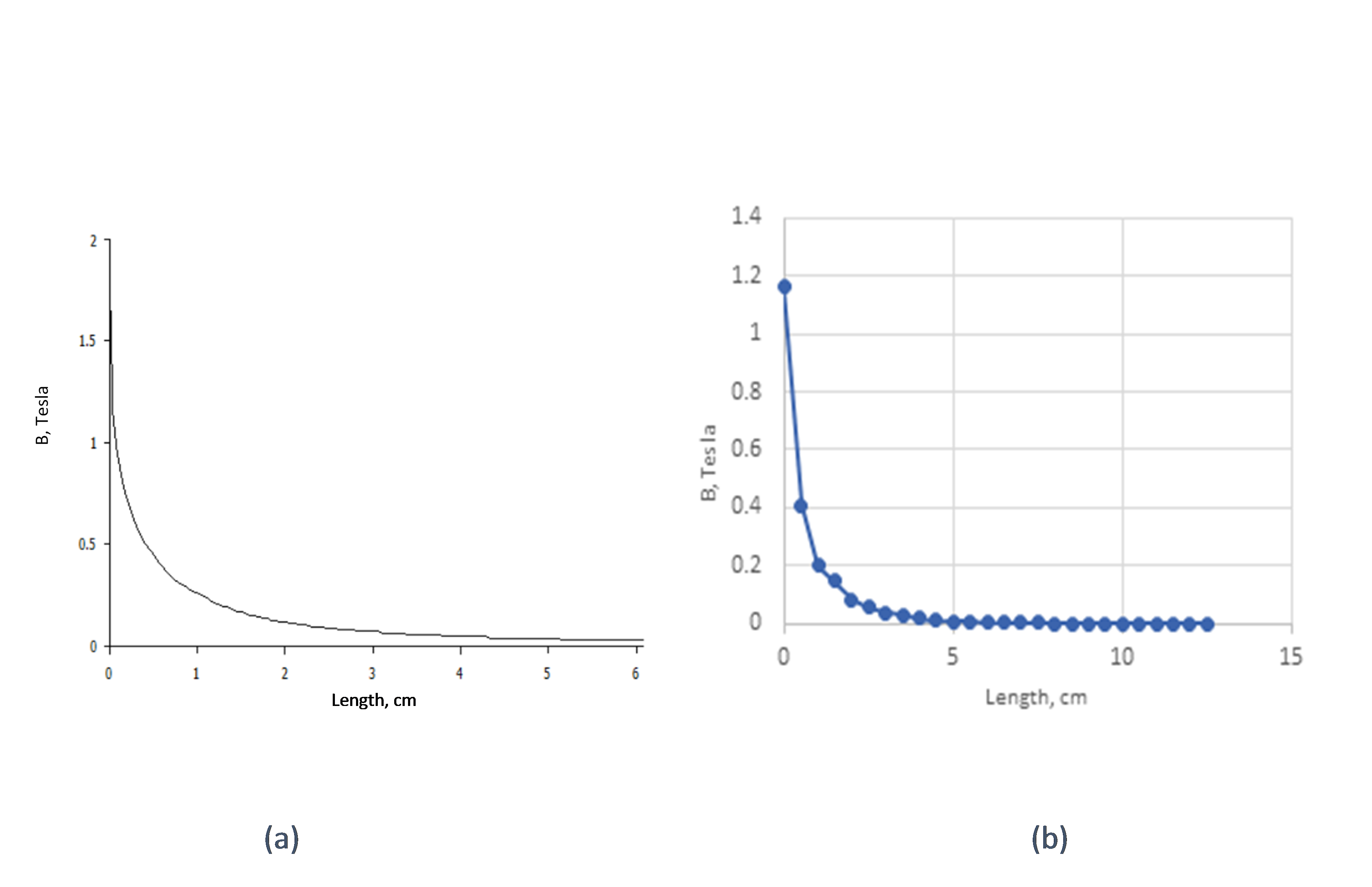
Computational model: A 2-D simulation model of a Neodymium-iron-boron (FeNdB-52) magnet was run using FEMM software. Halbach arrays were employed in the model design in order to maximize the strength of the magnetic field and field gradient. According to the FEMM model, the maximum strength of the Halbach array is 1.6 Tesla.
In vitro model: The FEMM model was assembled and actually produced a magnetic strength of 1.1 Tesla and magnetic field gradient of 37.4T/m up to 3 cm as well as a magnetic force of 43.4T2/m and 11.2T2/m up to 3cm and 12cm respectively.
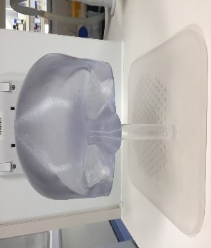
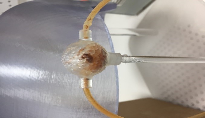
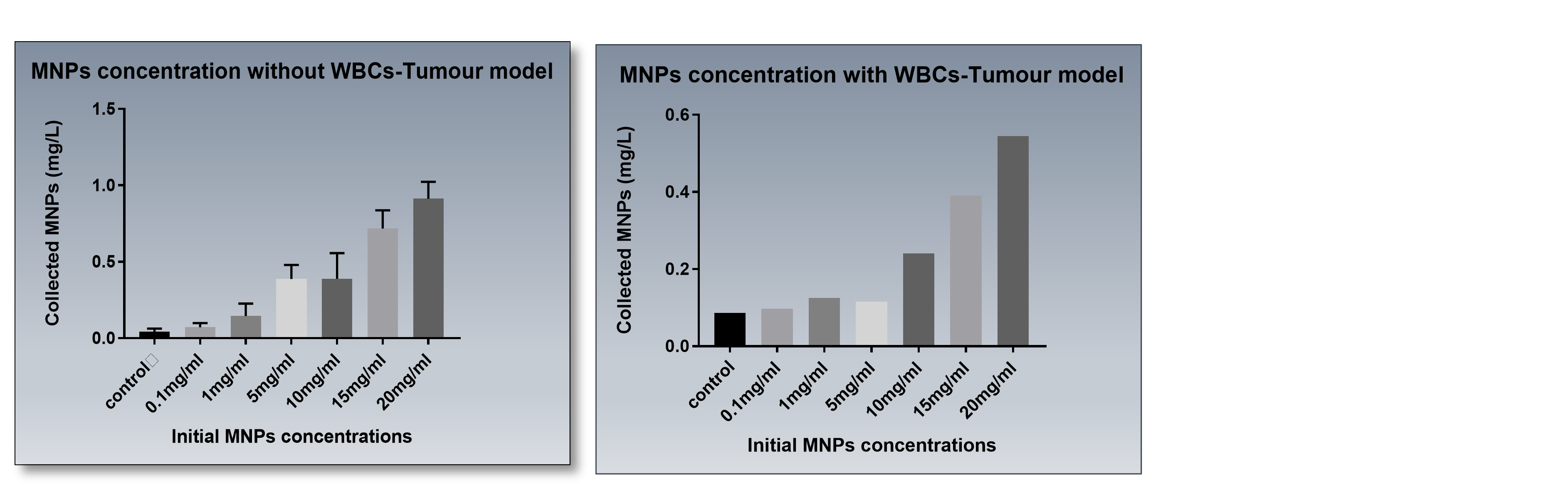
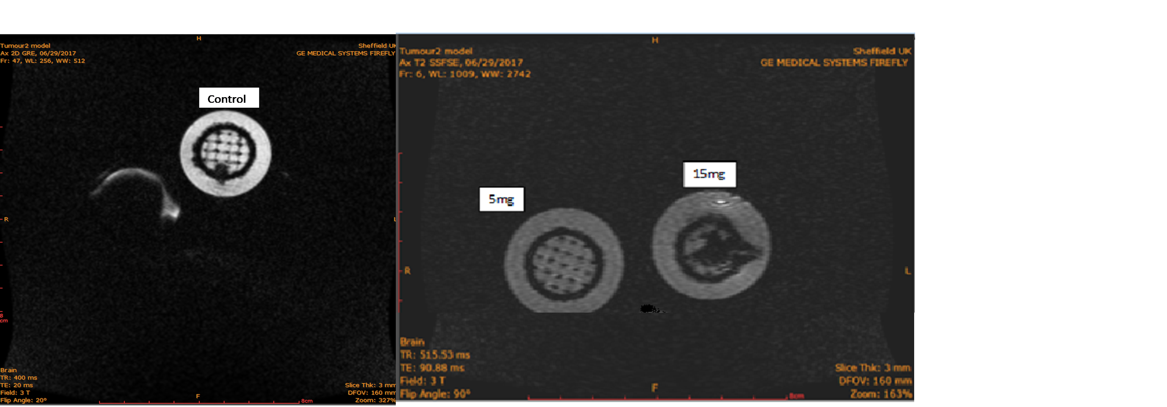
From optical and MR scan data, a 3D head/tumor model was printed which geometrically mimics the surface of the head of a patient. We also printed a brain tumor, with a positioning matrix allowing the tumor to be relocated anywhere within the printed head (Figure 1). A Halbach array magnet was placed on the top of the head in order to trap Fe3O4 MNPs in the tumor, in contact with the surface of the head model (0.8 cm thickness). MNPs were run through a fluid flow system using a syringe pump of 10 ml/min (Master Dual Pump,WPI.) Basically, the experiment was performed in two parts; the first part was to flow MNPs with different concentrations ranging from 0.1mg/ml to 20mg/ml. The second part was to flow MNPs, with the same concentrations, but in the presence of 50 million white blood cells (WBCs). After MNPs were added to WBCs, they were kept in an incubator (370c) overnight. The concentration of MNPs was quantified by inductively coupled plasma spectrometry (ICP). Images of the tumors with trapped MNPs were also obtained by MRI (3T) using a dual gradient echo sequence (TE= 4.60ms, 20ms).
Discussion and Conclusion
Various Halbach array designs were modelled and an optimised design assembled. The strength of the magnet measured using a Gauss meter was slightly less than calculated via FEMM (Figure 2). The in vitro experiments showed that Halbach arrays could trap MNPs w/o WBCs inside the tumor successfully (Figure 3). ICP data shown in Figure 4, demonstrated that, for 20 mg/ml of MNPs, 0.9 mg/l of MNPs flowed past the magnet as well as for 20 mg/ml of MNPs loaded with WBCs, 0.5 mg/l of MNPs/WBCs flowed past the magnet. From MRI it was clear that the rest were trapped in the tumor (Figure 5). Thus, ICP and MRI data confirmed the trapping ability of the optimised Halbach array. We are now focusing on trapping MNPs w/o WBCs into the model tumors over increased distances and with a more complex brain vascular network prior to developing an in vivo model.Acknowledgements
No acknowledgement found.References
Muthana M, et al (2015). Nat Comms 6:8009.Figures